According to an open-access article in ARRS’ American Journal of Roentgenology (AJR), the use of spectral CT with electron density imaging improved the assessment of lung lesion extent in patients with early-stage coronavirus disease (COVID-19).
“In the present study,” wrote Beatrice Daoud and colleagues at Antony’s Private Hospital in France, “we report the first retrospective data from the spectral chest CT findings of patients with reverse transcription–polymerase chain reaction (RT-PCR)–confirmed COVID-19 (i.e., patients with positive RT-PCR test results).”
Since March 17, 2020, every patient who has had CT performed at the authors’ institution for either suspected or RT-PCR–confirmed COVID-19 has undergone dual-layer detector–based spectral CT (IQon Spectral CT, Philips Healthcare). To evaluate the potential benefit of spectral imaging—electron density imaging, especially—two experienced thoracic radiologists reviewed the cases of four patients who each underwent two chest CT scans for confirmed COVID-19. Reconstructing the spectral CT images using the same standard soft kernel (filter B) and a similar iterative method that was used to acquire the conventional CT images, Daoud’s team also compared initial conventional CT images with follow-up conventional CT images.
In all four patients, their pulmonary lesions (45 ground-glass opacities, overall) were more conspicuous on electron density images than on initial conventional CT images and were clearly confirmed on follow-up conventional CT images. Moreover, lesion extent, assessed via semiquantitative reporting scale denoting surface area involvement for each lobe, was easier to ascertain on electron density images.
With Daoud and colleagues’ results indicating electron density imaging improves early assessment of the extent of ground-glass opacities that could be missed by conventional CT, electron density showed the most promising results by enhancing the contrast of ground-glass opacities compared with the normal lung.
“We reviewed conventional chest CT images obtained with a parenchyma kernel and standard lung window setting, as is usually the case in everyday radiology practice,” Daoud et al. explained, adding that they compared these images with conventional images obtained using a soft mediastinum kernel and standard lung window setting, conventional images obtained using a soft mediastinum kernel and narrow lung window setting, virtual low-monoenergy images, virtual high-monoenergy images, and electron density images.
“Our results suggest that the better ground-glass opacity visualization obtained using electron density imaging may be chiefly related to the increased visual noise in the image with soft kernel reconstruction and narrow lung window setting compared with electron density imaging, for which narrowing the window does not impair image quality,” the authors of this AJR article concluded.
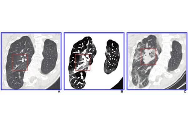
Featured image: 41-Year-Old Autistic Woman With Major Scoliosis and COVID-19: A, Initial conventional axial CT image shows no noticeable lung damage (within red box) in right upper lobe. B, Electron density spectral CT image obtained at same time as image in A shows lesions (within red box) in right upper lobe. C, Follow-up conventional axial chest CT image obtained 5 days after images in A and B confirm presence of lesions (within red box) in right upper lobe. (courtesy: AJR)


