 The theory that “men are from Mars, women are from Venus” applies to more than communication styles.
When diagnosing physical problems of the heart, the genders offer different symptoms. Men who experience heart attack, for example, typically have crushing chest pain or pain that radiates down the left arm and sometimes into the throat and jaw. Women communicate heart problems with what experts consider atypical signs and at an older age than men.
The ability to identify the signs and order the appropriate test to diagnose cardiac problems is crucial to saving lives. For years, men were the center of research and attention for cardiac disease. However, attention is shifting with the recognition that a heart attack is the No. 1 killer of men and women today, and it is just as important to diagnose cardiac problems early in women as in men.
While imaging modalities offer both men and women the promise of accurate diagnosis, treatment and cure, overcoming the challenges posed by a woman’s physical makeup remain a challenge for some modalities more than others. And while not all tests are alike, each specialty, predictably, will champion its own modality.
“Because women present with different symptoms, it is pretty hard as a doctor to know what to do,” says Rory Hachamovitch, M.D., co-director of nuclear cardiac imaging and director of cardiac SPECT (single photon emission computed tomography) at St. Francis Hospital (Roslyn, N.Y.). “That little bit of twinge they have in their chest that you can say with confidence doesn’t sound like angina in a man, but in a woman, you don’t know what to do. [The result] is that women are referred for noninvasive testing at much lower risk than men because of this uncertainty about symptoms.”
Depending on what kind of patients a physician sends for a test, how well the test will perform becomes very different. If the wrong population is sent, it will not perform as well as if the “correct” patients are tested. “This is something that has been long recognized and has driven how we decide which patient should go for which test,” Hachamovitch adds. “We don’t have a great way to predict risk in women, and it’s something that is being very actively worked on.”
Transthoracic echocardiography
Echocardiography, along with its imaging counterparts, is benefiting from emerging technology. Leonard Ginzton, M.D., professor of medicine at the University of California at Los Angeles (UCLA) and director of echocardiography at Harbor-UCLA Medical Center (Torrance, Calif.), says the failure rate (failing to allow visualization for a diagnosis) with echo at Harbor-UCLA is far less than 1 percent. “Harmonic imaging and the use of contrast agents markedly improve the image quality, and that will allow us to make measurements of quantitative blood flow, which are now not possible by any of the techniques, except possibly PET,” he says.
Approximately one-third of the patients undergoing an echo exam at Harbor-UCLA Medical Center are referred from its nuclear department, which does not use nuclear imaging for obese patients. The medical center also disallows nuclear tests on anyone with lung disease, referring them for echo as well, because pharmacologically induced stress can cause asthmatic attacks in that group.
Stress echocardiography is one of the most commonly used imaging modalities to detect coronary artery disease in women and men, but its critics say it can’t identify a low-risk patient population.
“The down side of stress echo is that you may be able to identify disease correctly, but if you have a normal stress nuclear study, the risk you have of an adverse event — meaning death or myocardial infarction — over the next year is in the range of 0.5 to 0.7 percent per year, the threshold that is accepted as low risk,” Hachamovitch says. “On the other hand, with stress echo, there’s only been one study to date that has a recorded event rate of less than 1 percent, and most of them have event rates of not only 1 percent, but of over 2 percent.”
Hachamovitch conducted a study with the Harvard School of Public Health (Boston), looking at all published papers on stress echo and stress nuclear cardiology. They studied data on patients who had suspected or known coronary disease and excluded anyone who recently had a heart attack or bypass surgery. The papers were based on up-to-date SPECT and echo techniques. They eliminated redundant papers (those published in multiple places by the same group) and focused on the event rate associated with a normal study. (Taken as a whole, a normal study is low risk.)
When Hachamovitch and his colleagues combined the papers and adjusted for the differences in population, they calculated how many people were going to die or have heart attacks in a population had a patient received stress echo instead of stress nuclear exams.
“It was clearly an excess number of extra events that occur, and you can quantitate how many extra events you would have missed because these people have gone to no extra treatment,” says Hachamovitch. “This is something that the echo community has to deal with. They can identify disease, but for some reason, they cannot identify a low-risk patient population.”
Transesophageal echocardiography
Because the images are so good, transesophageal echocardiography (TEE) has evolved for a number of indications. There is no evidence that the modality would be better for one gender than another. TEE can provide clear pictures of the heart for women or men who are very obese, have bad lung disease, have had recent chest surgery and can’t be rotated on their left side, and for people on respirators.
 |
 |
| An FDG PET scan found good viability throughout the myocardium, except for a small part of the apex, in this heart image of a 50-year-old female. System: Siemens Medical Systems’ ECAT PET scanner for all clinical applications. | |
“You can see things in areas of the heart and of the great vessels, especially the aorta, that aren’t visible from the outside with any transthoracic imaging,” explains Paul Tunick, M.D., cardiologist and professor of clinical medicine at New York University School of Medicine (New York City). “Or if they’re visible, they’re very poorly seen from transthoracic echo.”
The five-minute TEE test has more indications than originally intended. “The most important indication is in patients who have had a stroke,” adds Tunick. “This is something we are very interested in, but it hasn’t gained widespread acceptance yet, and it’s very important. You can identify the cause of stroke in a lot of patients where it’s impossible otherwise.”
Tunick says that for most people who have had a stroke, everyone thinks of carotid artery disease or atrial fibrillation, but there are just as many people with large plaques in the arch of the aorta that have caused the stroke. “We’re doing [transesophageal echo] in practically every stroke patient,” Tunick says.
The modality also is useful in patients with artificial heart valves, infections in the heart or congenital heart abnormalities. It’s also playing a much bigger role during heart surgery, helping prevent stroke from occurring, which can happen in as many as 5 percent of patients. By locating plaques in the aorta, the surgeon can avoid hitting them during the surgery.
Gated SPECT
Gated SPECT (single photon emission computed tomography) allows the nuclear cardiologist to obtain images of the heart at multiple points. The process includes gating certain windows of time during the cardiac cycle. The modality provides reproducible and quantifiable maps of blood flow at both rest and stress, as well as left ventricular volume and left ventricular ejection fraction (the percentage of blood the heart ejects with each beat) before or after stress.
At the very least, nuclear testing works as well in women as in men. The modality identifies low-risk women and men equally well, but relatively high-risk women more accurately than relatively high-risk men — despite the fact that the literature says nuclear testing doesn’t work for women.
“It becomes a function of how you do the test,” St. Francis’ Hachamovitch explains. “Today, the majority of [SPECT] studies are done with Technetium 99m Sestamibi, which is a brighter, hotter agent that gives us much better images.”
Sestamibi also allows cardiologists to measure both how well the heart is squeezing and the blood flow. Hachamovitch says a case is growing in literature showing that it is much more accurate to do testing in women with sestamibi than with thalium. “If you do it [with sestamibi], you’re going to get every bit as good a result as you’re going to get with other noninvasive tests,” he says.
Breast attenuation has created problems in obtaining accurate images with SPECT. But attenuation correction and nuclear cardiology expertise have limited the attenuation factor.
“Breast imaging artifact isn’t really an issue if you look at the really large trials which have looked at outcomes for women and men,” says Ronald Schwartz, M.D., director of nuclear cardiology at the University of Rochester Medical Center (Rochester, N.Y.). “Despite whatever relatively minor issues with lung vs. breast imaging artifact for echo vs. SPECT which exist, it’s very well known that SPECT perfusion imaging yields incremental prognostic value for both women and men.”
Women are substantially higher risk for problems like bleeding, infections, heart attack or stroke from invasive tests. “SPECT provides the risk information needed to be able to match the level of therapeutic intervention,” Schwartz says. “It does not give us the anatomic map we need. For that we need arteriography, if we decide the situation is risky enough to warrant it.”
EBCT
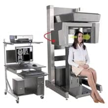
To some, electron beam computed tomography (EBCT), which identifies amounts of calcium in the heart, should be the modality of choice for diagnosing heart problems in women. Its clinical use is increasing, according to Matthew Budoff, M.D., assistant professor of medicine at the Harbor-UCLA Medical Center. “It’s actually better in women than it is in men,” Budoff says. “It’s more specific in women and is a better test [in women because] the electron beam enters through the back, so the radiation is in the back of the heart.”
Because the beam doesn’t have to go through the breast tissue to get to the heart, there is no problem with the breast obscuring the heart, and the breast is actually shielded from the radiation source. “Since most women’s backs are not as muscular as men’s, the images are even more clear in women than in men, which might be an advantage, and that would really be a difference from all the tests we have in cardiology today,” Budoff says. “When we talk about the possible advantage of any technology in dealing with women, you would want to go through the back, not have a lot of breast exposure and not be inhibited by breast tissue.”
The controversy with EBCT is in the asymptomatic patient, according to Budoff, in trying to take a person who has no symptoms who might be at risk for heart disease and screening an otherwise healthy person with the modality.
The American College of Cardiology (ACC of Reston, Va.) and the American Heart Association (AHA of Dallas) have prepared a statement suggesting that EBCT is not ready or recommended for routine use in patients.
According to the two organizations, a high calcium level in the heart indicates an increased risk of coronary disease, but it doesn’t necessarily mean that the patient has coronary disease. People with kidney disease, for example, get calcifications in their hearts whether they have coronary artery disease or not.
“If we were to do electron beam CT in that patient population, a high proportion would be positive, whether they have coronary artery disease or not,” says Myron Gerson, M.D., professor of medicine and radiology at the University of Cincinnati College of Medicine. “Essentially, it just would not answer the question. A lot of people will be identified as being at some increased risk, and they will need to go on to some further testing. That’s one of the reasons the ACC and AHA have released this position statement that electron beam CT is not ready for [routine use] yet.”
Positron emission tomography (PET)
PET is well beyond the research stage, but its disadvantages of relatively high test cost and the small number of experts in the modality have limited its growth. PET shows perfusion and wall motion. It images blood flow, as well as receptors and metabolism in any part of the body.
“PET can do things no other modality can ever imagine doing, and as we try to be more sophisticated in the questions we ask, PET is going to have a better and better future,” says Hachamovitch.
PET images metabolism by using the agent fluorodeoxyglucose (FDG), which is considered by most to be the standard for detecting viability — that is, what parts of the heart muscle are still alive and thus justify a patient’s having a revascularization.
Medical centers that use PET for myocardial imaging indicate the overall results compared to SPECT show it has an edge. One of the problems has been whether it is enough of an improvement to justify the increase in cost for doing PET.
“The gain does seem to be primarily in women and because of the accurate attenuation correction that can be done with PET compared to SPECT,” opines Edward Coleman, M.D., professor of radiology at Duke University Medical Center (Durham, N.C.). “So if one were going to take a group of patients who would benefit most from the PET scan compared to SPECT imaging, it would be women.” Coleman adds, however, that because the overall number of false positives in women is not that high in SPECT, that for a 5 to 10 percent gain, most have thought it was not worth the additional effort or expense to do PET instead.
 |
 |
| These cardiac SPECT images show a normal woman’s heart on the left and an abnormal heart on the right. The images represent three orthogonal SPECT projections of the heart — at stress on top and at rest on the bottom. | |
PET also faces the challenge of reimbursement for determining myocardial viability. Reimbursement is being considered by the Health Care Financing Administration (HCFA) and may be decided by June.
Utilization of PET overall is expected to increase. “Now that PET is being reimbursed in oncology, there will be a lot more PET scans out there, and I think we will see increasing utilization in evaluating the heart,” says Coleman.
Cardiac MRI
For evaluating the structure of the heart, most clinicians would argue that MRI is the gold standard. But the modality is considered by many to be not ready for clinical utility for general heart imaging. The heart has always been one of the most difficult parts of the body to image with MRI because of the motion of the heart. The issues of breathing and beating require MRI to be fast enough to be able to image the heart while it’s moving in order for the modality to be useful.
The technology to enable this is starting to get out to the community, according to Vivian Lee, M.D., Ph.D., assistant professor of radiology and director of cardiothoracic imaging at New York University (New York City). “Cardiac MR right now can look at cardiac function, left ventricular wall motion to look for areas that don’t work very well or areas that may be damaged from coronary artery disease,” says Lee. “It can be used to look at myocardial perfusion and to identify how much damaged or scarred heart tissue there is, including whether a patient has had a myocardial infarct in the past.” It also can look at myocardial masses, pericardial disease, as well as congenital heart disease and valve abnormalities.
MRI is only in its infancy, however, in visualizing the coronary arteries themselves. Lee says that many people are waiting for MRI to be able to visualize the coronary arteries because there’s a feeling that it should be a one-stop shop. “I disagree with that, but many people feel for MRI to be cost effective, it needs to be able to show the coronary arteries like an angiogram, in addition to doing perfusion and functional study,” says Lee.
Part of MRI’s role is to become a means to eliminate unnecessary coronary catheterizations. Lee is interested in female patients who may benefit from going directly to MRI. “Women patients may undergo an echocardiogram or a nuclear [cardiology] scan and the results will be equivocal, so the doctors then will send them on for coronary catheterization,” explains Lee. “There may be a disproportionately high number of patients who have to undergo coronary catheterization who have absolutely normal coronary arteries, and those are the patients we could possibly help out in MR.” More women than men have negative coronary catheterizations.
As research continues, the real cardiac imaging questions for the future, St. Francis’ Hachamovitch says, are: Is there any difference in modalities; what will it cost for this difference? And, how well can the average person do the test to interpret the information?
Today, most experts agree: The test women and men should choose for imaging the heart is the one that is performed expertly in a person’s community. ![]()


