Cancer treatment has evolved into three distinct approaches. The first and oldest is direct destruction of the cancer epicenter through surgery; the tumor and adjacent tumor-bearing areas are mechanically removed. The advantage of this approach is that the excised tumor cells are guaranteed to be destroyed. Removing an irregularly shaped, infiltrating mass from the patient without causing considerable collateral damage is not easy, however, and many ingenious operations have been developed to accomplish this task. Unfortunately, destroying the tumor’s home base is often insufficient to cure the patient, as malignancies can spread far from the original site.
Direct tumor destruction is also carried out through the use of x-rays. Radiation therapy has been used for cancer patients for more than 100 years. The technique works because cancer cells are more sensitive to the damaging effects of radiation than are normal cells. This makes it possible to destroy tumor cells that are intermixed with normal cells selectively. This is impossible with surgery, but radiation therapy has many of the same limitations as surgery: the radiation oncologist must know precisely where the tumor is located, and systemic treatment with radiation is not currently possible.
The second approach to treating cancer uses systemic chemotherapeutic agents. These chemicals can penetrate into most body compartments and kill cancer cells more readily than normal cells. This property seemingly relieves the treating physicians from having to know just where the cancer resides, but chemotherapy is often much less effective than it should be. For example, a large lung tumor may seem to shrink away completely after the first two cycles of multiagent chemotherapy, only to have it return after a few months. Often, the recurrent tumor will be unaffected by further chemotherapy, even if new drugs are used. This phenomenon, known as multidrug resistance, appears to be a fundamental, evolutionary capability of many mammalian cells that permits them to withstand environmental toxins. Resistance may always limit the value of conventional chemotherapy.
The third and newest approach to cancer therapy is the use of highly selective agents that target specific characteristics of tumor cells, such as antibodies to tumor-specific antigens and agents that target certain metabolic pathways. Because these agents are so specific, they are much less toxic than conventional chemotherapeutic agents. Whether these highly specific agents are curative is still unclear; tumor cells may find ways to resist them.
Of course, no one knows which, if any, of these three general approaches to cancer treatment can be significantly improved through further research. Perhaps the wisest strategy is to pursue all three.
The Role of Imaging
Radiation therapy relies on the fact that tumor cells are more easily destroyed by radiation than normal cells. If radiation is employed as a local-regional treatment (as is usual), then normal tissues outside the radiation ports can compensate and can partly replace those that are damaged or destroyed within the radiation ports. To achieve the delivery of high radiation doses to the tumor while limiting doses to nearby normal structures, one must use multiple radiation beams that overlap only on the tumor. To do this correctly requires two distinct technologies: one to locate the tumor accurately and the other to aim the radiation beams correctly.
Tumor location can sometimes be determined by palpation or inferred because of its particular disruptive effect. In the majority of cases, however, the location of the tumor must be determined using studies that either acquire an image of the tumor directly or infer its probable location from surrounding normal anatomy. Sometimes, both approaches are needed.
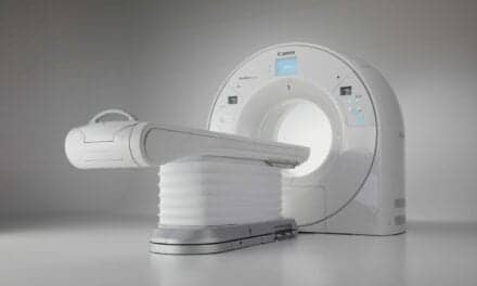
In the past, radiation oncologists had to rely on planar radiographs, which were rarely of value in determining gross tumor volume (GTV), except in the lung. Currently, the most common imaging used for radiation therapy is CT, from which GTV can be determined either because the tumor’s radiographic density differs from that of surrounding normal tissue or because the tumor distorts or destroys that anatomy. CT scans have been most valuable in the lung, mediastinum, and pelvis.
MRI differentiates tumors from normal tissue because water content is frequently greater in tumors than in normal tissue. MRI has been most useful in delineating GTV in the brain, head, neck, abdomen (especially the liver), and extremities. Magnetic resonance spectroscopy imaging (MRSI) extends the capability of MRI by imaging differences in the chemical composition of tissues, not just their water content. Proponents of MRSI hope that, in time, they will be able to determine unique tumor-chemical signatures that will provide the clinician with more information about the tumor than just its location.
A third approach to tumor imaging is functional imaging, which relies on metabolic differences between tumors and normal tissues. The most familiar functional imaging is probably the bone scan, wherein osteoblasts in the bone take up a disproportionate amount of technetium. A gamma camera images the technetium uptake, showing areas of abnormal bone metabolism that can, given certain clinical circumstances, indicate bony metastatic lesions. Similarly, other agents can be used to image specific tumors.
Currently, the most interesting approach to tumor imaging is positron-emission tomography (PET) scanning. The most commonly used PET agent is fluorodeoxyglucose (FDG), which is taken up by tumor and normal cells along with ordinary glucose. FDG can be made with an isotope of fluorine that emits a positron (the antimatter version of an electron). The emitted positron will usually travel less than a millimeter before interacting with an electron. The result of contact between a positron and electron is the annihilation of both particles and the production of a pair of 511-keV photons, or gamma rays, that flee the scene in opposite directions. A pair of waiting cameras can easily detect the highly specific 511-keV photons, so PET scanning can give a three-dimensional (3D) map of glucose uptake in the imaged region. Since tumors are usually far more metabolically active than normal cells, PET scans can be highly effective in distinguishing tumors from normal tissues, even when CT or MRI scans are normal. Combination PET and CT scanners can provide simultaneous metabolic and anatomic information about a patient. Studies of the value of PET/CT scanners in radiation treatment planning will begin shortly in a number of centers.
Even the best CT or MRI scanners cannot detect tumors much less than 2 mm in size, and PET and MRSI fail at about 8 mm. It is just as important to treat unseen, microscopic disease as gross disease, so the clinical tumor volume (CTV) must still be determined using knowledge of the pathways through which tumors spread. Fortunately, CT and MRI scans are helpful in explicitly demonstrating the surrounding normal anatomy, which is crucial to guiding the clinician in determining a reasonable CTV.
Treatment Planning
Identifying the GTV and inferring the CTV are only the first steps in planning. This information must be used by 3D treatment-planning software to guide the radiation beams. The patient is first CT scanned under controlled conditions, frequently using the same immobilizing device in which he or she will be treated. A fiducial mark is placed on the device or patient so that accurate correspondence can later be made between the scan and the patient. The diagnostic scans used to define the GTV and CTV are then spatially registered with the planning CT scan. Both manual and automatic methods for performing this registration have been developed, and these methods are gradually finding their way into commercial treatment-planning software.
The next step is the most difficult and important. The clinician must manually outline the GTV and CTV, as well as certain important normal tissues and organs. Errors in defining the CTV may well result in failure to cure the patient, whereas incorrect identification of normal tissue structures can result in increased morbidity. Dividing a medical image into sensible anatomic regions is image segmentation. Automatic segmentation has been the goal of many in the field of image processing. Unfortunately, image segmentation seems to require more than just determination of changes in radiographic density between one structure and another. Even well-defined objects, such as the kidneys, have boundaries that are often indistinct or missing. Software that tracks these boundaries will fail.
Good planning software presents the segmented image data in a variety of formats suitable for designing radiation beams. One of the most successful of these is the beam’s-eye view, which shows a digitally reconstructed radiograph (DRR) from the viewpoint of a candidate radiation beam. These DRRs look much like ordinary radiographs, but can have the segmented data superimposed on them. This enables the clinician to design radiation beams that fully encompass known and suspected tumor sites. DRRs can also, through comparison with port films, be used to see whether a radiation beam is being correctly delivered. In prostate and lung cancer, use of 3D planning appears to be improving cure rates; in head and neck cancer, it seems to be reducing treatment-related morbidity.
The response to chemotherapy can be quite variable, even within a single patient. According to current thinking, treatment with chemotherapy should continue at least as long as there is measurable tumor response, with the assumption being that responses seen in the GTV predict responses in the part of the tumor that is too small for imaging. Once the tumor becomes drug resistant, chemotherapy can only be harmful to the patient, so methods for precise tracking of the GTV can provide a rational stopping point for chemotherapy.
Systemic Disease, Local Therapy
What might be accomplished if a highly tumor-specific scan could be performed with a spatial resolution of 100 ?m? Mammography can already achieve this goal because microcalcifications are so much brighter than the surrounding tissues. The average human cell is approximately 10 ?m long, so a densely packed sphere of 1,000 malignant cells would have a diameter of about 100 ?m, making it detectable by the hypothetical scanner. What would a whole body scan of a lung-cancer patient with supposedly localized disease show? Because we now know that many of these patients die of systemic disease, even if their local disease is removed, it is clear that these patients must have micrometastatic disease somewhere in their bodies at the time of presentation. At the 100-?m level, would the disease be manifested as a diffuse process throughout the entire body or as a countable number of nodules that might be amenable to individual (local) treatment?
The ratio of patients presenting with a small number of metastatic sites to patients with an uncountable number of sites (diffuse disease) is largely unknown, but we do know that at least a few patients must present with a limited number of metastatic sites. This inference can be drawn from studies of patients who present with solitary metastases. In many malignancies, 20% of such patients can be cured by surgical excision of the metastatic site (metastasectomy). Patients can even be cured of multiple solitary nodules of colon cancer metastatic to the liver and, perhaps, a few other diseases. Therefore, it is reasonable to suppose that an imaging device able to detect 100- ?m tumors could identify patients with metastatic disease who could be cured using local modalities.
Even more intriguing would be the use of such a device for patients with more diffuse metastatic disease. Patients with small amounts of diffuse metastatic disease can be cured using chemotherapy; this is the rationale for adjuvant chemotherapy in breast, lung, and colon cancer. If patients have enough metastatic disease to be imaged using current techniques, however, they are rarely curable using chemotherapy. The difference must lie in the amount of metastatic disease present. Perhaps high-quality imaging would allow physicians to render the patient curable through chemotherapy by showing them where the larger metastatic deposits lie, thus permitting their destruction using local modalities.
Julian Rosenman, MD, is an oncologist in the Department of Radiation Oncology, the University of North Carolina School of Medicine, Chapel Hill, NC


