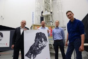
From left, Randolf Hanke, Michael Böhnel, Nils Reims, and Anne Schulp. Photo courtesy Fraunhofer IIS/Peter Roggenthin.
The Development Center for X-Ray Technology, part of the Fraunhofer Institute for Integrated Circuits IIS, recently presented exclusive computer tomography (CT) images of a Tyrannosaurus rex.
Describing it as one of the best preserved T. rex finds of all time, researchers from the Naturalis Biodiversity Center in Leiden, Netherlands made the discovery in Montana. Experts date the remains of the female dinosaur to 66.4 million years. The skull alone weighs 500 kilograms.
“This discovery will have an enormous impact on dinosaur research for decades to come,” said Edwin van Huis, head of the Naturalis Biodiversity Center.
To get a glimpse of the internal structures of the remains without damaging the fragile skeleton, researchers utilized one-of-a-kind XXL CT technology from the Fraunhofer Development Center for X-Ray Technology in Fürth, Germany.
“We’re extremely pleased that the Naturalis Biodiversity Center has placed their trust in us,” said Randolf Hanke, head of the center. “With its unique CT technology, Fraunhofer’s Development Center for X-Ray Technology can make a significant contribution to help shape dinosaur research.”

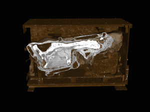
The images are computer tomography scans of a major Tyrannosaurus rex skull taken at the Fraunhofer Development Center for X-Ray Technology. Photo courtesy Naturalis Biodiversity Center/Fraunhofer IIS.
The precise, cross-sectional x-rays of the skull benefit the conservation and preservation of the remains. Surprises, such as hidden fractures, can be reliably detected in advance and then taken into consideration during the preparation. Furthermore, using the x-ray data and a 3D printing process, a true-to-original copy of the skeleton can be produced.
“Concealed areas are especially interesting for us,” said Anne Schulp, paleontologist and dinosaur researcher at the Naturalis Biodiversity Center. “With this method, we are in a position to reconstruct the structure of the skeleton, especially since the skull is in such excellent condition in this case. I’m extremely excited about the mold of the inside of the skull. We can show what the brain looked like without having to open up the irrecoverable skull.”
Get AXIS e-newsletters free. Subscribe here.


