From the Experts: Ultrasound Present and Future
Ultrasound and Women’s Health
Niche Player Now Fills Multiple Needs
SonoSite Brings Customization to Women’s Health
 |
From the Experts: Ultrasound Present and Future
Axis Imaging News recently spoke with leading manufacturers in the field of ultrasound about current trends and future prospects. The participants included: Terri Bresenham, vice president and general manager of Global Diagnostic Ultrasound for GE Healthcare; Klaus Hambuechen, CEO, Ultrasound, Siemens Healthcare; Anne LeGrand, senior vice president and general manager, ultrasound, for Philips Healthcare; Gordon Parhar, director, Ultrasound Business Unit, Toshiba America Medical Systems; and Lars Shaw, vice president of marketing, ZONARE Medical Systems.
IE: What are some of the trends that we’re seeing in ultrasound? What are the demands of customers these days?
 |
| Terri Bresenham |
Hambuechen: One of the major trends in ultrasound is certainly the ever-increasing information density to let physicians achieve higher diagnostic confidence. At the same time, this goes hand in hand with the demand for decreased study time. This seems to be a contradiction. Increased information density can easily lead to information overflow. And what good is an increased amount of data if medical professionals can’t make use of it? Siemens’ ACUSON ultrasound systems feature intelligent, knowledge-based algorithms that automate and streamline a broad range of routine clinical tasks. Using learned pattern recognition from an expert database of thousands of real clinical cases, knowledge-based workflow applications recognize anatomical patterns and landmarks and can automatically perform measurements for a whole new level of accuracy and efficiency. Knowledge-based workflow technology enables clinicians to achieve consistent and reproducible measurements with high speed and fidelity across a wide range of applications and users. Examples of knowledge-based workflow applications are, among others, syngo® eSieCalcs™ native tracing software, which features proprietary border detection technology to facilitate lesion or anatomical structure boundary segmentation, and syngo AutoOB measurements, which allow automatic biometric measurements of the fetus (BPD, HC, AC, FL, HL, CRL). Another trend along these lines is 3D/4D imaging, which provides additional diagnostic information. The technique was predominantly used for fetal imaging, eg, for the diagnosis of facial anomalies, especially facial clefts, but is evolving into other areas such as general imaging and cardiology.
Shaw: We are seeing a much greater demand for portable radiology ultrasound studies. In fact, clinicians tell us they need premium image quality and features to be brought to the bedside, as these patients are the most difficult and complex exams they do. However, space is very limited, so the conventional large refrigerator-type systems are not suited for this new type of ultrasound study. Ease of use, boot-up time, battery-powered, cordless scanning, and a full range of transducers for any type of exam they might encounter are all needs that are growing very fast because of the need for portable ultrasound exams. The hand-carried or compact ultrasound business is also growing rapidly. Because a compact ultrasound unit can fit into many different spaces, several different departments within the hospital and private offices are also creating demand for both inpatient and outpatient needs. For example, interventional radiology, vascular labs, emergency departments, urology, ob/gyn, cardiology, and more. Even though they each have different clinical ultrasound imaging needs, they all share a common need as well. They require a premium-performance, high-quality diagnostic imaging system that is easy to use and portable and works efficiently.
 |
| Anne LeGrand |
Parhar: Ergonomics continues to be of focus as ultrasound systems become smaller and lighter. Miniaturization and improvements to current control panel designs make systems more user-friendly and customizable and prevent work-related musculoskeletal disorders (WRMSD). Another major focus is to improve diagnostic confidence through improvements in image quality by using new signal processing techniques and transducer designs, and by making the exam more reproducible and less user dependent. Customers are demanding innovation along with exceptional value. Value is a key driver, especially in the current economic environment.
LeGrand: We are seeing an increasing demand for ultrasound exams and the need to do more exams per day, per machine. There is also a demand for more diagnostic ultrasound information to increase confidence when radiologists are making the call. In addition, there is an increasing demand for an ultrasound solution for obese and technically difficult patients.
There is a rising expectation of greater ultrasound performance and feature sets for the money, especially for mid-range products. Also, customers expect vendors to make ultrasound system optimization easier with fewer keystrokes. Finally, customers want greater performance for compact products to maximize diagnostic confidence at the bedside. Beyond radiologists, cardiologists, obstetricians, and gynecologists, ultrasound is being used by new classes of specialists, including anesthesiologists and emergency care physicians, even for clinics and hospitals in rural areas and developing countries. Improving performance of these systems is a key factor in driving use and demand, particularly in the operating room and critical care units, where space and budgets are limited and high-quality images are critical.
 |
| Klaus Hambuechen |
Bresenham: Ultrasound procedures are continuing to grow, most rapidly in areas like vascular, urology, and breast and in nontraditional segments such as anesthesia, vein access, ER, surgery, etc. There is an overall trend to portability as ultrasound procedures move closer to the patient’s bedside, coupled with a need for adept information technology to easily transfer ultrasound information around in this more distributed model. We also see a demand for ultrasound technology that can image more challenging and obese patients; penetration capability is becoming a differentiator for purchasers of ultrasound equipment. We also see growth in ultrasound’s role in interventional procedures. For example, new advances in volumetric image fusion technology, where live ultrasound can be fused in real time with CT, enable more procedures to be performed under ultrasound guidance. This benefits patients and clinicians by minimizing radiation dose, can improve overall workflow productivity, and often leads to better asset utilization for CT.
IE: What are you concentrating on in terms of future development? Where is the market headed and what steps are you taking to meet these emerging needs?
Bresenham: As one of the largest providers of ultrasound technology, GE is very positive about the ways ultrasound can help health care meet its critical needs—lowering costs, providing ubiquitous access, improving the quality of care, reducing radiation exposure, and getting real-time information in a caregiver’s hands. We also believe that ultrasound has a long runway left before realizing its full potential as a diagnostic imaging technique. For instance, ultrasound is one of the highest resolution imaging techniques, but in a conventional system, scatter detracts from the inherent exquisite detail. With 12 R&D centers globally, we are actively engaged with health care systems around the world to take advantage of this inherent but unrealized opportunity. Our primary technology investments are centered around one goal: Solve clinically relevant problems. With so many ways for ultrasound to benefit patient care in a cost-effective manner, in many ways, it feels like we just got started.
 |
| Gordon Parhar |
LeGrand: A big part of the future of ultrasound for radiology is volume imaging—allowing radiologists access to a block of ultrasound information similar to the approach they use for CT and MR. All of the pieces are starting to come together to make this transition easy for radiologists. Philips’ unique xMATRIX technology will provide a one-transducer solution for outstanding 2D, 3D, Live Volume, and Live xPlane imaging performance. In the area of volume workflow, PACS will have the DICOM standard to retrieve ultrasound volumes and workstations will fully integrate a host of automated tools that make volume imaging easy and quantitative. New volume analysis tools also provide the ability to examine serialized views of the area or lesion over time—a significant development for therapy monitoring.
New technologies that offer more diagnostic information will continue to play a major role in the evolution of ultrasound. We expect to see more developments in image fusion of live ultrasound with CT/MR and other modalities. Also, an easier, but more advanced, approach to elastography that makes it reproducible and quantitative and continued breakthroughs in ultrasound image quality. As in the past, these advanced technologies will migrate from the premium systems to the mid-range and compact products.
Parhar: We are focused on clinical excellence and will continue to explore and develop new methods and technologies that provide faster, more accurate diagnosis and a better patient experience. Health care cost-containment concerns will continue to plague the diagnostic medical imaging community. Ultrasound is best positioned as a cost-effective modality, and Toshiba is continuing to make the necessary investments to ensure we have the necessary solutions as ultrasound becomes the preferred modality and its use becomes more widespread in the United States.
 |
| Lars Shaw |
Hambuechen: Siemens is continually developing new technologies to push the envelope of clinical workflow through innovations. We are working to improve imaging performance and create new applications. Going forward, we are exploring the possibilities of a generalized scanning robot for different applications and of arrays that can scan a large area without moving the transducer at all. For new applications and imaging performance, we are also looking into the image fusion of ultrasound and CT for interventional guidance.
Silicon Ultrasound is one of our most important areas of development, offering a whole new level of imaging performance that will ultimately be integrated into our S Class premier imaging platforms. Silicon Ultrasound technology introduces the first entirely new class of ultrasound transducers in 40 years. It uses the precise semiconductor processing techniques of the computer chip industry to create a family of probes that will enable volumetric 4D imaging in a wide range of applications. With Silicon Ultrasound, clinicians will get true isotropic 3D images, enabling them to see the same fine level of detail in each direction that they choose to examine the imaging data. Siemens demonstrated the power of this approach in breast imaging and other high-resolution applications at RSNA 2008. (NOTE: Robot, image fusion of ultrasound and CT, and Silicon Ultrasound are not currently commercially available in the United States.)
Shaw: At ZONARE, we continue to advance our image quality, performance, and clinical features to bring premium performance to wherever clinicians need diagnostic confidence in their ultrasound study.
 |
Ultrasound and Women’s Health
On a clear day, a pilot in an airplane is able to see all the peaks and valleys below. Fly into a thunderstorm, and the pilot won’t be able to see anything.
This example is how Erin Owens, clinical marketing senior manager for the ultrasound business unit of Toshiba America Medical Systems, explained the company’s new MicroPure technology for ultrasound-guided stereotactic biopsy. Showcased at the recent RSNA meeting, MicroPure essentially removes the thunderstorm from the picture by placing microfilm over a B-mode image and bringing out calcifications.
“In breast imaging, one of the hardest things to see is microcalcification, and ultrasound has a very difficult time seeing these, especially 700 microns and lower,” Owens said. “What we’ve done is developed a technique to bring these calcifications out so we can better see them.”
According to Toshiba, its new MicroPure technology aids physicians in the detection of microcalcifications using ultrasound, which is less strenuous on the technician and patient than mammography. Owens notes that ultrasound will not replace mammography, but the imaging technique would be performed on patients with suspected calcifications. MicroPure involves a one-touch button that allows doctors to identify calcifications quickly, putting less strain on anxious patients undergoing biopsy.
 |
| MicroPure technology aids physicians in the detection of microcalcifications using ultrasound. |
Also at RSNA, Toshiba unveiled its work-in-progress ElastoQ technology, a quantification of the company’s elastography package. Like MicroPure, the noninvasive imaging technique is not meant to replace something that already exists. Instead, it was developed as another tool to use in diagnosis, evaluating tumors based on stiffness compared to normal tissue.
According to Toshiba, the company’s method is highly reproducible. Tissue is evaluated while it is slightly decompressed and compressed, as cancerous tissue tends to exhibit significantly lower elasticity than healthy tissue.
Currently, ElastoQ is undergoing clinical evaluation at Northwestern University in Chicago. Owens reports that while the company has not received all the results just yet, it has seen that in the preliminary stages, the technology is comparing very closely to BIRADS.
Owens also said the technologies received substantial positive feedback at the Chicago radiology meeting. Attendees were particularly interested in what further techniques they could be used with—for MicroPure, thyroid and musculoskeletal imaging; for ElastoQ, going beyond breast imaging and into prostate applications.
“Increasing the ability to use ultrasound to image the breast is a significant development,” said Gordon Parhar, director, Ultrasound Business Unit, Toshiba. “We believe these advances will benefit many women across the country.”
Both technologies will be available on Toshiba’s Aplio XG ultrasound.
Niche Player Now Fills Multiple Needs
With the advancements that ZONARE Medical Systems Inc has made to its ultrasound platform in just 1 year, the Mountain View, Calif, developer aims to compete with the big boys.
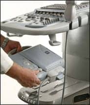 |
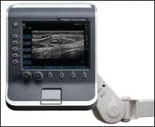
| ZONARE recently introduced several advancements for its z.one ultrasound system, a premium-quality convertible ultrasound platform. |
“Instead of just being that niche player of just portables, we’ve gotten feedback that we can fill complete radiology department needs with our new releases,” said Lars Shaw, vice president of marketing.
The releases to which Shaw was referring were all shown at the Radiological Society of North America meeting. At its booth, ZONARE unveiled an echocardiography release for the company’s z.one ultra system, which can now offer premium image quality and advanced color and spectral Doppler sensitivity, as well as new TEE capabilities and a comprehensive calculations package for left ventricle analysis and valvular pathologies.
“Last year, we showed basic elementary cardiac images,” Shaw said. “Now, we have an offering that can do full cardiography exams bedside or in the department.”
Commenting on the echo technology, ZONARE president and CEO Don Southard said the solution helps address the critical needs of busy clinical practices.
“By bringing the premium performance echocardiography release to the z.one ultra system, we are expanding the value and versatility of our customers’ investment in ZONARE,” Southard said. “This release represents all the elements of a complete premium performance solution.”
ZONARE also displayed software advancements to its proprietary Zone Sonography technology, which, when combined with new transducer technology, makes up a new package for difficult-to-image patients. “Most companies, when they sell you their system, the instrument is as good as it’s ever going to get,” Shaw said, adding that most conventional ultrasound companies make upgrades to hardware, a costly and lengthy process. ZONARE’s advancements, on the other hand, are completely software based. “When you buy our instrument, it just gets better and better all along,” Shaw continued.
For elderly, obese, or muscular people, or those with thick body walls, the C4-1 curved array transducer allows for improved penetration with sensitive Doppler imaging and a small footprint.
The company also showcased a new L14-5w high-resolution, high-frequency transducer. Through its broad bandwidth, the product can better image small parts, breasts, and superficial anatomy. Ten frequencies include three fundamental, one tissue harmonic, two compound imaging, and two each for color Doppler and PW Doppler modes.
Additionally, the z.one ultrasound system can come equipped with new elastography applications, which enable qualitative visual assessment of the mechanical stiffness properties of tissue. Using a variety of grayscale and colorized maps, high-resolution elastography images are visualized.
Comprehensive 3D ultrasound imaging functionality is available with primary applications for obstetrical imaging during the second and third trimester, as well as for general abdominal ultrasound imaging.
SonoSite Brings Customization to Women’s Health
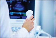
One fell swoop of the hand is all it takes for women’s health clinicians at the Center for Breast Care in Burbank, Calif, to transport SonoSite Inc’s latest ultrasound technology from one room to the other.
 |
| The S-Women’s Health ultrasound tool from SonoSite enables physicians to address a range of women’s health concerns. |
“The S-Women’s Health is great to have in the exam room because if a patient comes in with a lump, I am able to quickly and easily determine if it is something like fibrocystic thickening, or if it requires additional evaluations,” said Deanna J. Attai, MD, FACS. “When you consider the quality and affordability of the system and the fact that you can pick it up and take it to the operating room, you just can’t get a better tool for your practice than the S-Women’s Health System.”
The S-Women’s Health ultrasound tool is the newest product in the Bothell, Wash, company’s S Series product line. Developed with the needs of physicians in mind, the custom-designed product joins the M-OB/Gyn Office and the S-Gyn ultrasound tools as part of SonoSite’s comprehensive solution for women’s health.
Engineers have omitted the bundle of buttons, dials, and menus from the interface and instead created two controls: Depth and Gain. Touted by the company as being sophisticated and simple, these controls are responsible for the acquisition of optimum image quality. Additionally, the development team has incorporated the company’s proprietary advanced technologies—SonoHD and SonoMB—which allow for clear imaging and precise visualization. SonoAdapt allows clinicians to focus on a targeted area, permitting users to accurately “locate masses, target lesions, guide biopsies, and initiate a course of treatment,” according to the company.
Sporting an award-winning design, the S-Women’s Health tool can be placed on a stand or mounted on an exam room ceiling or wall. Enabling physicians to address a range of women’s health concerns, the system is configured to work with four broadband transducers to provide high-resolution superficial, abdominal, vascular, and pelvic imaging.
Moreover, the company offers a 5-year warranty, which covers the system and transducers. This warranty allows Attai to do her job with the secure knowledge that if the product malfunctions, her patients’ care will not be compromised.
“Not only does SonoSite have superior products, but they also have superior service,” Attai said. “The 5-year warranty on their products means that not only will they repair the system if needed, they will get a loaner to me the very next morning. I can’t practice without my ultrasound system, and SonoSite recognizes that.”
This latest product announcement comes after the release of the S-Gyn during the Annual Meeting of the Radiological Society of North America, which took place in Chicago in late November into early December. Also custom-designed, the S-Gyn was streamlined for gynecologists to perform imaging for diagnosis and procedural guidance in the patient exam room.
“I have two great big machines in my office, but for a lot of what I’m doing in my day-to-day practice, such as evaluating abnormal bleeding, checking the ovaries or uterine structures for polyps or fibroids—I can do all that with the S-Gyn without having to move my patient out of the exam room,” said Shaunie Keys, MD, Evergreen Women’s Care, Kirkland, Wash. “A big advantage of the S-Gyn system is its portability and that I don’t have to disrupt workflow by shuffling patients to different rooms or interrupt my ultrasound tech’s schedule.”
Describing additional benefits of the S-Gyn, Keys commented, “It’s easy to learn and provides really good imaging. My patients feel more confident knowing that I will complete the ultrasound exam and they will have their results before leaving the office.”
Like the S-Women’s Health ultrasound, the S-Gyn is equipped with SonoHD, SonoMB, and SonoAdapt, and it comes with the company’s 5-year warranty. Furthermore, the product’s small footprint allows it to fit easily beside the patient exam table or mounted to the ceiling or wall.
The S-Gyn is configured to use the C60x abdominal and ICTx transvaginal transducers.
SonoSite offers training and education programs that can be delivered on site, over the Internet, and in collaboration with medical institutions and educators.
Other products in the S Series family of systems include the S-Nerve for anesthesiologists, the S-Cath for interventionalists, the S-ICU for intensivists, the S-Fast for emergency medicine physicians, and S-MSK for musculoskeletal specialists.