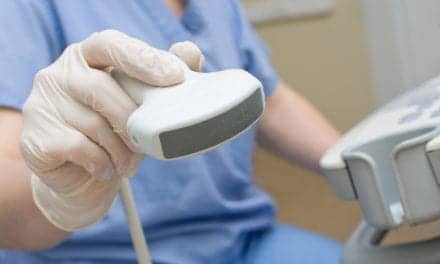
The Aixplorer® Ultrasound System with ShearWave™ Elastography allows clinicians to determine quantitative liver stiffness values in a non-invasive, easy-to-use exam, which can be safely repeated over time to follow disease progression or regression.
Makers of Aixplorer®, SuperSonic Imagine’s revolutionary technology is now being applied to liver imaging, possibly reducing the need for many biopsies. Liver biopsy has traditionally been considered the standard for assessing liver fibrosis severity but this invasive method has major drawbacks including significant incidence of morbidity, procedure and hospitalization costs, and clinical shortcomings as fibrosis is underestimated in 10-30% of the cases. In addition, because of the invasive nature, biopsy is not ideal for repeated follow-up exams.
ShearWave Elastography is a non-invasive technique that is being used worldwide to visualize and quantitatively measure (in kilopascals) tissue stiffness across the different stages of fibrosis leading up to cirrhosis. This diagnostic information can trigger medical treatment, help to evaluate the progress and effectiveness of drug therapy, and provide regular and previously unavailable imaging monitoring for complications. When invasive procedures are called for, Aixplorer’s exceptional image quality has proven highly effective in helping hepatologists and radiologists with ultrasound guided liver procedures such as needle placement for biopsy and paracentesis.
According to SuperSonic Imagine CEO Jacques Souquet Ph.D. “Several clinical studies have concluded that ShearWave Elastography is an accurate, reproducible technique to assess liver disease. The impact of ShearWave™ Elastography in liver imaging, both in clinical and economic terms, cannot be underestimated. This technology will enable a major shift in patient management.”


