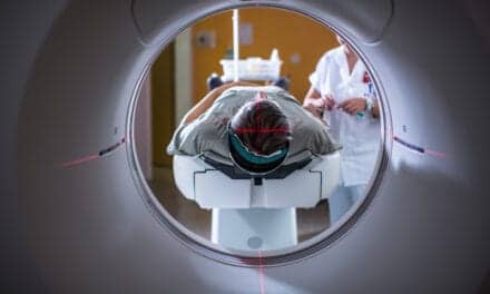
Researchers at Imperial College London have developed a prototype of a mini MRI scanner that fits around a patient’s leg. The team members say the device—which uses a so-called ‘magic angle’ effect—could potentially help diagnose knee injuries more quickly and accurately.
In a study using animal knees, the results suggest the technology could be used to show all the structures of the knee. The scientists say the device, which looks like a large metal ring through which a patient places their leg, could help diagnose conditions such as anterior cruciate ligament injuries—which are particularly common among footballers. Furthermore, the small size of the device could enable it to be used in local clinics and even GP surgeries, potentially reducing NHS waiting times for MRI scans.
Currently, key components of the knee joints such as ligaments and tendons are difficult to see in detail in the MRI scans, explains Karyn Chappell, PhD, a researcher and radiographer from Imperial’s MSK Lab: “Knee injuries affect millions of people and MRI scans are crucial to diagnosing the problem, leading to quick and effective treatment. However, we currently face two problems: connective tissue in the knee is unclear on MRI scans, and people are waiting a long time for a scan.”
Chappell adds: “This can cause particular problems for women, as they are at greater risk of anterior cruciate ligament injuries. The reasons for this are unclear, but it could be linked to hormones such as estrogen making ligaments more elastic, leading to more joint injuries.”
Knee injuries commonly affect one of three areas: the tendons, the meniscus, or the ligaments. However, tendons, ligaments, and meniscus are not usually visible with MRI, due to the way water molecules are arranged in these structures, explains Chappell.
She adds: “These structures are normally black on an MRI scan—they simply don’t produce much signal that can be detected by the machine to create the image. This is because they are made mostly of the protein collagen, arranged as fibers. The collagen fibers hold water molecules in a tight configuration, and it is in fact water that is detected by the MRI. If you do see a signal it suggests there is more fluid in the area—which suggests damage, but it is very difficult for medical staff to conclusively say if there is injury.”
To overcome this problem, Chappell harnessed the power of a phenomenon called the ‘magic angle’: “The brightness of these tissues such as tendons and ligaments in MRI images strongly depends on the angle between the collagen fibers and the magnetic field of the scanner. If this angle is 55 degrees the image can be very bright, but for other angles it is usually very dark.”
The team explains that the magic angle is achieved in their scanner because they are able to easily change the orientation of the magnetic field. While the patient sits comfortably in a chair, the specially designed magnet, which uses motors and sensors similar to those found in robots in car factories, can rotate around the leg and the orientate magnetic field in multiple directions.
This is not possible in current hospital MRI scanners, which are also much more expensive than the prototype scanner.
“Previously the magic angle phenomenon was thought of as a problem, as it could mean medical staff mistakenly thinking the knee is injured. However, I realized that if we took a number of scans around the knee, we could use the signal produced by the magic angle effect to build a clear picture of the knee structures,” explains Chappell.
“Specifically, we can combine images obtained at different magnet angles and not only increase the brightness, but also see how the collagen fibers are arranged. This enables us to establish the pattern of collagen fibers in the knee structures, which is crucial information ahead of treatments such as repairing a torn meniscus,” adds Chappell. “At the moment, it’s very difficult to see which direction the collagen fibers run in a meniscus. This is important because sewing across the fibers will effectively repair a tear in the meniscus. However, if the stitch is in the same direction as the fibers, the repair may fail.”
In a new study, published in the journal Magnetic Resonance in Medicine, the multi-disciplinary team scanned the knee joints of six goats and ten dogs in a conventional MRI scanner. All of the dog legs were donated by the Royal Veterinary College, having been donated for research by dog owners following the death of their pet. Dogs suffer from knee injuries and arthritis similar to humans, making them a good subject for the study.
The results showed that using the magic angle can accurately detect ligament and tendon damage. The team say now they know magic angle scanning can be used to visualize the knee, combining this with the new prototype mini scanner could enable knees to be accurately scanned with this technology and hope to progress to human trials of the ‘mini’ scanner within a year.
Chappell explains: “Although this is an early-stage proof-of-concept study, it shows the technology could potentially be used to accurately detect knee injury. We now hope to enter human trials and explore if this technology could be used for other joints such as ankles, wrists and elbows.”