The medical community is waiting for the results of CT colonography studies that are under way before deciding to recommend and reimburse the modality as a screening method for colorectal cancer. Supporters think this is just a formality.
From its inception, CT colonography (CTC) has held great promise. Use of the imaging technology to examine the colon reduces the discomfort of the exam, eliminates the need for anesthesia, and has zero risk of perforating the bowel. The less invasive exam is not as intimidating to patients. As a result, supporters predict that the CT option will encourage previously wary patients to agree to regular colorectal cancer screenings and therefore decrease related mortality.
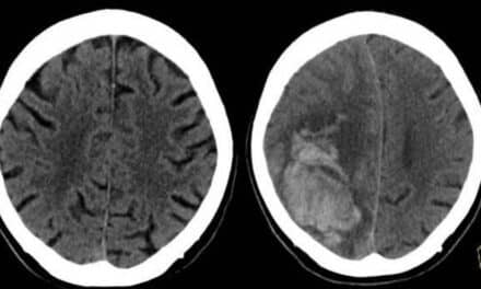
According to the CDC, colorectal cancer is one of the most commonly diagnosed cancers in the United States. In 2003, 73,182 men and 70,763 women were diagnosed with the disease; 27,990 men and 27,793 women died as a result. These related deaths, however, could be reduced by as much as 60% with regular screening.
-

- This system lets the user know which portion of the colon has not been visualized.

Currently, the CDC supports guidelines that suggest patients with normal risk begin regular screening at 50 years of age; those at higher risk can start earlier. Recommended tests include an annual fecal occult blood test (FOBT), flexible sigmoidoscopy every 5 years, double-contrast barium enema every 5 years, and a colonoscopy every 10 years.
For some patients, however, a colonoscopy even once a decade is too often; the CDC estimates that more than 60% of those older than 50 years have not been screened appropriately. CTC supporters suggest that if the newer method were recommended and reimbursed for screening, many more patients would submit to examinations according to the guidelines, reducing the impact of the disease over time. According to the CDC, when colorectal cancer is found early and treated, the 5-year relative survival rate is 90%.
-

- Vendors are making advances in technology as the field expands.

But these numbers are not what is necessary to convince the medical community of the modality’s value—actual evidence of the procedure’s benefits as a screening method is needed to win over both practitioners and payors. This is rarely accomplished overnight; large multicenter trials take time, and the process has been hindered by studies with conflicting results. Some research has shown CTC’s sensitivity to be comparable to that of colonoscopy; others have shown it to be far less. Explanations point to the level of technology and the experience of the readers, so more studies have been conducted to mimic clinical environments. One of the largest, and perhaps the most eagerly anticipated, studies under way is that of the American College of Radiology Imaging Network (ACRIN).
The organization has completed collecting data for a study comparing CTC and standard diagnostic procedures in detecting colorectal neoplasia in 90 patients. It has also recruited and gathered data from more than 2,500 healthy patients to examine CTC as a screening tool for colorectal cancer. Experts expect the results will be published within the next year.
“The last major step [to CTC acceptance] is publication of the ACRIN study. Does this mean that if the results suggest high sensitivity, 30 days later CTC will be reimbursed? No, the wheels will have to be in motion, but a lot seems to be waiting on these trials,” said Zaffar Hayat, president of Viatronix Inc, Stony Brook, NY.
Waiting on Reimbursement
Much of what is holding up widespread adoption of CTC as a screening method is reimbursement. “Reimbursement is a critical step for CTC screening to really take off,” said Perry J. Pickhardt, MD, associate professor of radiology in the abdominal imaging section of the University of Wisconsin School of Medicine, Madison. He attributes the lack of reimbursement to the confusion regarding mixed performance results.
“[These results] can easily be explained by technical differences, for example, 2D versus 3D polyp detection,” Pickhardt said. He was the lead author on a 2003 study that found CTC had sensitivity for the detection of polyps comparable to that of colonoscopy and in some cases better. The research employed advanced technology and experienced readers, which some argued did not represent a realistic clinical environment.
As technology and training have become more common, however, it is less unreasonable to expect these factors to be achieved in today’s clinical facilities. Current studies have often incorporated the latest advances.
Because the highest quality technology and readers are clinical standards at the University of Wisconsin School of Medicine, the facility was able to use its research data to negotiate reimbursement from local HMOs for CTC as a screening method. “It is unreasonable to expect folks to pay out of pocket, even for a potentially preferred test like CT,” said Pickhardt.
“A lot of progress is being made month by month in getting local coverage decisions to cover CT colonography for specific indications like incomplete colonoscopy or patient refusal due to reasons such as anticoagulation medication,” said Ronald M. Summers, MD, PhD, tenured senior investigator and staff radiologist at the National Institutes of Health Clinical Center in Bethesda, Md. He feels this can be expected to expand if the ACRIN study reports positive results.
Waiting on Accuracy
Many expect, or at least hope, that the ACRIN results will duplicate the capabilities seen in the Pickhardt and other similar studies and lead to further reimbursement. “By that, we mean sensitivities in the eighties or greater for polyps 1 cm or larger and a specificity of at least 90%,” said Summers. Polyps 10 mm or larger are generally referred for a polypectomy. Those smaller than 5 mm are typically left alone. Pickhardt said, “Large polyps—10 mm and greater—are the target for colorectal screening with CTC, since they have the greatest risk of becoming cancer. Diminutive lesions—5 mm and less—are not clinically relevant for CTC and should not be reported.”
Medium-size polyps, those in the 6 mm to 9 mm range, are more controversial. Pickhardt suggests they may not always be a danger. “For polyps 6 mm to 9 mm detected by CTC, it is quite possible that the risks and costs associated with polypectomy outweigh the tiny potential risk of future cancer from a small lesion,” said Pickhardt.
A study published in April 2007, for which Pickhardt was an author, found that medium-size lesions were more likely to have a low prevalence of high-grade dysplasia and invasive carcinoma. Of course, the medical community wants more data, but if the evidence holds up, physicians may decide to recommend surveillance rather than removal for medium-size polyps without other suspicious qualities. “Generally, polyps grow about 1 mm per year, but it takes time to build a database,” said Viatronix’s Hayat.
“If [medium-size polyps aren’t always removed], fewer colonoscopies will have to be scheduled following CT colonography,” said Summers, a fact that could alleviate some logistical challenges. Same-day removal is preferred by many facilities and patients, but the uncertainty can make effective use of resources difficult. Subsequently, some polypectomies will be scheduled at a later date, a process that may be inconvenient and/or inefficient.
Waiting on Tools
CTC is more sensitive with larger polyps, although shape may also impact accuracy. “Flat polyps are said to be the hardest to find with CT colonography, but there is evidence that they don’t occur very often,” said Summers. Other factors to monitor include bowel distension, bowel prep, lack of contrast, and reader training. According to Summers, these can result in false negatives; false positives may result from residual stool or inadequate distension.
To reduce issues with residual stool or fluid, contrast agents are becoming a standard part of the procedure; indeed, they have been a tool since CTC’s inception. “I believe that oral contrast tagging—preferably with both 2% barium and diatrizoate—is now the standard of care for CTC and should be used whenever possible,” Pickhardt said.
The tags permit electronic cleansing of the colon and are particularly invaluable when it cannot be cleaned. Summers notes that there have been a number of studies on the topic. “My view, based on my understanding of these studies, is that is important to have oral contrast to tag residual stool,” he said.
As the contrast agents become more effective, they may replace other, more involved preparatory methods and be used to actually “clean” the colon, reducing the need for extensive bowel preparation. Summers recommends iodine-containing contrast agents for tagging residual fluid (“although some can lead to diarrhea,” he warns) and barium tags for residual stool. In addition, he stated, “There is some evidence that using 3D endoluminal images leads to improved polyp detection, but most experts in the field think you have to use both 3D and 2D images to make the diagnosis.”
Waiting on Training
To do this accurately, though, requires training. Researchers are examining exactly how much is necessary, but no one questions that a certain amount should be required. “I’ve heard that 75 to 100 cases should be required,” said Summers.
-

- A translucent view of a polyp is shown in this image from Viatronix.

-
- Multiple views of the colon enhance the reading experience.

“A few studies have shown that if the radiologist is not well trained, performance is poor,” Summers said. He cites a study conducted in South Carolina that found reader accuracy ranged from 30% in one facility to 80% in another. A study published in the May issue of Clinical Radiology by Burling and others found that individual accuracy ranged from 53% to 93%.
The ACRIN study incorporates advanced technology and training qualifications. “The ACRIN trial required radiologists to undergo training and pass a test. Only those who passed were allowed to participate,” said Summers.
“Training is important for accurate interpretation,” said Pickhardt. In addition to being able to discern polyps, the reader needs to know how to use the technology. Pickhardt said, “Fortunately, the learning curve for the 3D approach is much simpler than for the earlier 2D search method.”
Viatronix’s Hayat notes that training opportunities have increased as the procedure has gained more support. “The purpose is not to push the platform but to teach participants how to more accurately diagnose,” said Hayat. Because the technology is new, many physicians did not learn it in school or even residency, and are uncertain which tools may be best for certain conditions. Though that is changing, associations, vendors, and individual groups, such as the University of Wisconsin, are organizing workshops.
Training, however, does not represent the hurdle that reimbursement does. “Lack of training is not as much of a barrier as the lack of reimbursement. Training can be accomplished rapidly,” said Hayat. “I’m sure that if CT colonography becomes reimbursable, there will be widespread interest by patients and physicians in the test, and people will take the training courses,” predicted Summers. But before that happens, the ACRIN results will need to be published and will need to be positive. Some see this as a foregone conclusion.
“Current state-of-the art CTC, including 3D polyp detection and oral contrast tagging, is at least as sensitive as the purported ‘gold standard’ optical colonoscopy for clinically relevant polyps. In fact, if done right, I believe it is the most sensitive test for detecting advanced neoplasia,” said Pickhardt.
For CTC supporters, the ACRIN study is merely a formality, necessary to bring the rest of the medical community on board. The buzz is already there. “During a meeting at the National Cancer Institute in early April, the agenda was centered around CTC and its adoption. It wasn’t so much about whether it’s a valid technique but how to move forward with it,” said Hayat. No one expects to take a step back, but they are all waiting to see.
Renee DiIulio is a contributing writer for Axis Imaging News. For more information, contact .
Real Tools for Virtual Reality
-

- New technology tools enhance viewing, accuracy, and efficiency.

As the medical community has debated the value of CT colonography as a viable screening method and study results have been published, vendors and users have proceeded to advance the technology, creating new tools to help manage the CTC application. Higher CT slices and the use of contrast tagging and electronic cleansing are examples that have already become virtually standard.
Newer technologies include flattening techniques, hybrid PET exams, and CAD, which is perhaps the furthest along with systems already in commercial use. Not everyone is sold on the value of these tools, however; Perry J. Pickhardt, MD, associate professor of radiology in the abdominal imaging section of the University of Wisconsin School of Medicine, Madison, said, “Because CTC is already so sensitive in its current state-of-the-art 3D form, it is unclear if any of the new technologies will play a major role in the future. Currently, they add little to the clinical evaluation.”
Others disagree. Ingo Schmuecking, MD, senior director marketing, image and knowledge management division, CAD and knowledge solutions, Siemens Medical Solutions, Malvern, Pa, believes that CAD tools can help to increase accuracy and efficiency. CAD can act as a second reader, pointing out polyps for investigation.
Siemens’ syngo Colonography with PEV (Polyp Enhanced Viewing) has been designed to detect polyps in the 6 mm to 20 mm range. “We have more than 107,000 CT exams in our database, which helps us very much with protocols. We started with clean prepped exams and are now working on tagged protocols,” Schmuecking said. The company seeks to design a database that is reflective of the clinical settings where the tools are used, developing technology to optimize subgroups such as flat or pedunculated polyps.
The system also offers tools for efficiency, such as auto-polyp measurement, which automatically measures the polyp, and auto-processing, which prepares the images for viewing by the radiologist automatically.
V3D-Colon from Viatronix Inc, Stony Brook, NY, also processes exams automatically. “The system removes other organs and sets up the flythrough in about 3 to 5 minutes,” said Zaffar Hayat, Viatronix’s president. Additional tools include automatic and interactive navigation, 100% lumen coverage and verification (achieved through different views), measurement tools, a transparent rendering mode, and 3D and 2D synchronized views.
—R. DiIulio