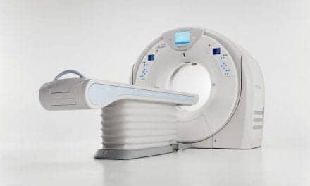
 The days of diagnostic x-ray angiography may be beginning to wane as less invasive modalities, such as CTA (computed tomography angiography) and MRA (magnetic resonance angiography), strut their technological stuff. While x-ray angiography, generally considered the gold standard, feels the competitive heat from diagnostic capabilities offered by CTA and MRA, the still-evolving angiographic players are not without remaining issues. Their absence is conspicuous in the interventional arena where x-ray angiography continues to wow the crowds.
As all three modalities wage the war against vascular disease — alone or as complements to one another — angiography is the vascular imaging workhorse when it comes to imaging diseased vessels from head to toe.
Certainly the statistics on diagnostic x-ray angiography indicate that the modality is following the trend for less invasive testing — the numbers for this application are down. However, just as Mark Twain remarked, “The reports of my death are a great exaggeration,” the same probably can be said for diagnostic x-ray angiography.
“I think the utilization of x-ray angiography has changed dramatically in the last five or so years because of advances, specifically in MR angiography and also CT angiography,” Neil Khilnani, M.D., associate professor of clinical radiology at the Weill Cornell Medical Center and New York Presbyterian Hospital (New York City). “There are certain areas where almost exclusively the diagnosis can only be gathered by x-ray angiography … There are certain cases when angiography is going to be essential for diagnosis, particularly when you get to smaller vessels. In children and in adults when you’re looking for small vessel disease, catheter angiography is staying [and] will probably stay for the foreseeable future.”
Where catheter angiography will persist is in the hands of people who aren’t just diagnostic angiographers or diagnostic radiologists, but rather interventional radiologists. Khilnani sees a clear and substantial shift at his institution from catheter-based angiography to MRA. “We are probably doing eight to 10 MRAs of the abdomen, pelvis and lower extremities per week, and in our outpatient facility we’re probably doing 25 a week.” That doesn’t include the MRAs that are done of the brain, which are also changing how carotid disease is worked and how intercranial vascular abnormalities, such as aneurysms, arterial venous malformations (AVMs) and some tumors are imaged.
“There’s no hard and fast rule which is the best or which one everything is going to go [to],” says Adam Hecht, M.D., a neurointerventionalist at Overlook Hospital (Summit, N.J.). Hecht’s work includes the embolization of intercranial aneurysms, occluding vessels or the actual diseased area. Using 3D angiography, the images are rotated in an infinite number of positions. An aneurysm can occur along many different areas of a particular vessel and may very well occur in an area from which another vessel branches.
Please refer to the November 2002 issue for the complete story. For information on article reprints, contact Martin St. Denis


