Could breast tomosynthesis replace digital mammography as the primary breast cancer screening tool? Should it? Will it?
Breast tomosynthesis could be the next big thing in breast cancer diagnostics and care—”could” being the operative word. The new modality has the potential to dramatically alter the mammography landscape in the United States. But before any transformation can happen, the first breast tomosynthesis system will need to be approved by the FDA.
Commercial availability in the United States could happen within the next year. Bedford, Mass-based Hologic Inc has already submitted an application for a breast tomosynthesis system to the FDA. Other manufacturers are expected to do so within the next couple of years.
If research provides the support needed for approval as well as clinical and commercial viability, tomosynthesis could possibly replace mammography as a primary screening tool for breast cancer. Early studies suggest the new modality could improve both the false-positive and false-negative rates that have limited the value of mammography.
 |
| Tubulolobular adenocarcinoma, not seen well in a conventional mammogram (left image), is much better appreciated in the slice from the tomosynthesis data set (right image). |
“[Replacement technologies] are usually flawed in one of many ways, for example, cost or sensitivity to cancer. Tomosynthesis is different. It really has pretty much all the advantages of mammography while addressing mammography’s Achilles heel,” said Joseph Lo, PhD, assistant professor of radiology and biomedical engineering at Duke University Medical Center in Durham, NC.
If breast tomosynthesis systems prove themselves in a clinical setting, they will offer a viable alternative to mammography, but the questions are many. “There are easily 20 or more unresolved questions,” Lo said. Unknowns include technique optimization methods, the role of CAD (computer-aided diagnosis), and the modality’s specific uses. Keys to adoption, such as reimbursement, workflow timing, and real-life statistics, are also unknown—for now. Clinical trials and early adopters (once the modality is approved) will help to work out some of these remaining issues.
In the meantime, the medical community and patients anticipate the modality’s arrival in the United States as a diagnostic tool. Lo receives about a call per week from a patient seeking entry into a clinical trial. “Word has been getting out from clinical trials suggesting that tomosynthesis is the answer” for women who recognize mammography’s limitations and/or have questions unanswered by mammography exams, according to Lo.
“Many of us are very excited, but by the same token we are to be scientific in how we study these technologies and their impact on patients, physicians, and insurers, if they do go forward,” said Mark A. Helvie, MD, professor of radiology and director of breast imaging at the University of Michigan Health System, Ann Arbor. For now, use remains investigational only.
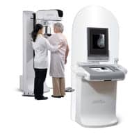 |
| Hologic’s Selenia Dimensions Breast Tomosynthesis System is awaiting FDA clearance. |
Benefits Suggest It Could
Breast tomosynthesis is essentially three-dimensional digital mammography. Using doses similar to those used in two-dimensional breast imaging, the system acquires multiple images of a stationary compressed breast from different angles. The images are reconstructed into high-resolution 1 mm slices that provide a three-dimensional view of the breast.
The slices reduce the noise typical of 2D images and separate the overlapping structures that could obstruct or mimic a lesion. This limitation has meant that traditional digital mammography had high false-negative and false-positive rates that got higher with denser breast tissue.
Mammography’s false-negative rates have been found to be about 20% and false positives 12%.1 Those figures change with patient demographics. Smith et al note that mammography detects approximately 90% of tumors in women over 50, but only 60% of tumors in women younger than that.1 Some suspect the numbers are even higher.
“In reality, [the false-negative rate] could be as high as 50% because the tumor remains small, lurks behind some other normal tissue, and simply isn’t seen,” Lo said.
With a 3D image, these problems are reduced. “The cancer that was hidden stands out in its whole slice,” said Lo, who compares the improvement to that seen in CT images over x-rays.
The technique is still not perfect, but even an incremental improvement is significant. “Most studies in the United States and Europe have shown incremental detection,” Helvie said.
The clearer view not only identifies lesions that may have previously been hidden but provides enough detail to allow better characterization. “If you can’t see a lesion in mammography, you can’t put a biopsy needle into it,” Lo said. But tomosynthesis may help patients to avoid the needle altogether.
False alarms are a huge drain on the resources of the health care institution as well as the patients. “It’s a huge waste of health care dollars, so being able to cut those costs by one-third or one-half would mean a substantial improvement in how we do breast cancer screening,” Lo said.
These savings are not a given, however. Although early research has shown that false-positive rates decrease with the use of breast tomosynthesis, actual use may bear out less rosy numbers. In a clinical situation, the intensity with which clinicians pursue a mass may be greater than in a research situation. “A 1% or 2% risk in research may not bother you, but in a clinical situation, you may be required to further evaluate that lesion,” Helvie said.
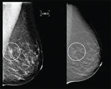 |
| Suspicious areas in a 2D mammogram (left image) can be resolved with 3D tomosynthesis (right image). |
Approval Decides Whether It Should
Similarly, a few more seconds in workflow may not seem like much, but in the clinical setting, already burdened with tight budgets and staff, the extra time can defeat a new technology. Naturally, systems’ timing will vary by manufacturer and improve over time.
However, the estimates sound workable. Hologic has suggested that its system takes a few seconds to acquire the image and a few more to reconstruct. “This would not produce a dramatic change in workflow,” Lo said.
Different techniques may take different amounts of time, particularly the more complicated algorithms, which require more processing power. “As research techniques prove themselves worthy, we would turn attention to optimizing them and speeding them up,” Lo said.
Once breast tomosynthesis systems are approved and in more widespread use, their true clinical value will be determined. “Most of us feel it is only a matter of time,” Lo said, noting that systems are already available in Europe.
Renee Diiulio is a contributing writer for Axis Imaging News.
Reference
- Smith AP, Hall PA, Marcello DM. Emerging technologies in breast cancer detection. Radiol Manage. 2004;26(4):16-24.