 |
Several of the factors that have facilitated the clinical adoption of positron-emission tomography (PET) in recent years continue to drive its spread today. One of these is the improving regional distribution of fluorodeoxyglucose (FDG), which makes it possible to provide PET scanning without owning and operating a cyclotron (which can cost $1.3 million to $1.8 million to acquire and a considerable additional sum to operate and staff) to produce FDG. Radiopharmaceuticals listed in the US Pharmacopeia already have the equivalent of approval by the US Food and Drug Administration, and FDG, the radiopharmaceutical with the longest half-life, is now available in many areas at a cost of $350 to $750 per dose, excluding delivery fees.
Another factor favoring PET’s dissemination is the influence of mobile PET services, which have convinced many referring physicians of the modality’s usefulness and prompted some of them to pursue PET acquisition for their own institutions.
SPACE REQUIREMENTS
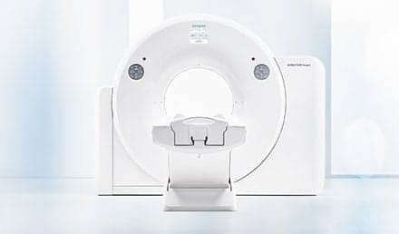
A PET system consists of the circular gantry into which the patient bed slides, the crystals arranged in rings that detect the annihilation g rays emitted by the radiopharmaceutical dose administered to the patient, the associated electronic circuitry, the lasers used to position the patient, the detector assembly, coincidence circuits, and the computer system that creates images from the scanner’s data.
PET centers should be designed to allow space for one or more scanner rooms, a control room, an injection room for each scanner room, a hot (radiopharmaceutical) laboratory, a utility room, a reading room, a workspace for nuclear medicine technologists (with suitable areas for patient monitoring), and a restroom area that includes a shielded hot toilet.
While it now seems possible for some PET providers to break even by performing as few as three PET studies or five PET-CT studies per day, siting should be planned with comfortable accommodation of maximal patient throughput in mind. This will vary by institution and scanner type, but can be expected to range from eight to 16 patients per day, per scanner.
The PET system’s manufacturer and type will dictate the amount of space needed not only for the scanner room, but for the control room and utility room associated with it. Minimum scanner-room dimensions vary from 3.9×5.8 m to 5×7.6 m for dedicated PET scanners and from 3.9×7 m to 5×7.6 m for PET-CT units. For dedicated PET scanners, control rooms’ sizes most likely range from 3.2×5.1 m to 3.4×3.8 m and utility rooms must be at least 1.8×1.8 m (and may need to be as large as 2.1×3.4 m). PET-CT scanners require control rooms of 2.5×3.9 m to 3.2×5 m and utility rooms of 1.8×1.8 m to 1.3×3.9 m. Each manufacturer will state the dimensions recommended for its system, but buyers may be able to make existing spaces that are not ideally shaped work for PET by exploring a variety of equipment placements. In particular, considering both centered and corner scanner placements may help PET planners use available space well.
The hot laboratory where radiopharmaceuticals will be handled should be arranged and situated in a way that minimizes handling times for radiopharmaceuticals; this improves imaging efficiency by taking better advantage of the short periods during which these agents are usable as tracers for imaging. A well counter for wipe tests, dose calibrator, sharps disposal for needles, and L-block shield should be available.
SHIELDING AND COOLING
The space required for PET scanning is specified by manufacturers as 39 m 2 to 58 m 2 , within a typical PET suite of 372 m 2 . The largest amounts of shielding will be needed for the injection room, hot laboratory, and PET-CT scanning room.
The hot laboratory should be shielded for 511 keV. This may require building alterations to support the necessary weight of shielding; for example, a lead L-block that is 5 cm thick weighs 272 kg. The hot laboratory will also need a source-storage/dose-storage area and syringe shields, along with a unit-dose shield weighing 30 to 33 kg.
The PET area will need sensitive temperature control, with high-velocity air conditioning and the ability to shut down the system automatically to protect the equipment if the specified temperature range is exceeded. A heat exchange/cooling loop will be required, in addition to a refrigeration system.
NETWORK AND POWER NEEDS
In choosing a location for PET scanning, planners must remember the need for connectivity. If this is overlooked, the organization may find itself required to make expensive modifications in order to extend its information network into the shielded space. The PET system’s computer should have access to the radiology information system, to the hospital information system (and electronic medical record, if in use), and to the picture archiving and communications system. Even if the enterprise has not yet implemented all of these information systems, future access to them from the PET suite should be prepared for during PET installation in order to avoid later disruption of operations.
The electricity to be made available to the PET suite must be specified during site planning; extra power may be required to meet the demands of the x-ray generator if a PET-CT system is to be installed.
PATIENT AND STAFF COMFORT
An injection room, where radiopharmaceuticals can be administered to patients prior to PET scanning, will be required. In order to minimize anomalous uptake of the tracer, it is necessary to keep the patient as quiet as possible. The sense of calm that is needed for this purpose can be promoted by ensuring that the injection room is free of all unnecessary, intrusive audible and visible stimuli. The number of patients that this room should be able to accommodate will depend on the uptake times of the radiopharmaceuticals used for PET scanning and the typical scan’s duration. Determining the probable waiting time and patient throughput for the PET scanner should lead to a reasonable estimate of the number of patients who will be waiting at any time.
The reading room for PET scans should have workstations for soft-copy viewing and may, depending on the preferences of the radiologists involved, need light boxes for hard-copy reading as well.
CONCLUSION
In the United States, procedural volumes for clinical PET appear to be increasing by approximately one third each year. This rapid growth is likely to continue as the areas served by air and ground deliveries from radiopharmaceutical suppliers expand. Hospitals of all sizes and types now offer PET services; attention to the modality’s siting requirements during planning will allow any institution to join their ranks without incurring unnecessary expense or unexpected delay.
This article has been excerpted from PET Imaging, which the author presented at the meeting of the American Healthcare Radiology Administrators, on June 28, 2002, in New Orleans, La.
Gary Sayed, PhD, is professor of radiological sciences, Charles Drew University of Medicine & Science, Los Angeles.


