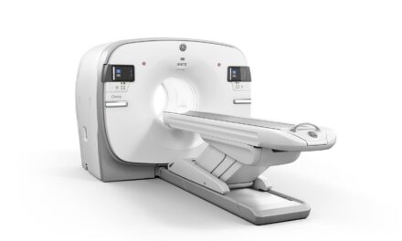
Sweden-based medical university Karolinska Institutet and MedTechLabs recently started testing a silicon-based, photon-counting CT system, which is designed for future clinical use, as they begin a new pilot study of the technology. The GE Healthcare device includes the company’s patented Deep Silicon detectors for photon-counting CT, which has the potential to deliver advanced spatial resolution without compromising count rate or spectral resolution.
News of this new clinical evaluation comes nearly one year after GE Healthcare’s acquisition of Prismatic Sensors AB, a Swedish start-up specializing in silicon detectors for photon-counting CT. Unlike other photon-counting CT detector materials, silicon has numerous advantages, including its purity, abundance, and broad manufacturing infrastructure, according to GE officials.
Historically, the main challenge with the use of silicon as a detector material is that it has a relatively low atomic number such that when placed in a “face on” position, it is too thin to stop and collect enough x-ray photons. However, using an approach that positions the silicon sensors “edge on,” GE Healthcare’s Deep Silicon detectors—which are made of pure silicon—can handle the very high photon flux (quantity of information) from the CT’s x-ray tubes. These silicon sensors absorb high-energy photons fast enough to count hundreds of millions of CT photons per second, which can create crisp images.
“While the system’s cover may look familiar, its potential capabilities are totally different—enabling us to image small blood vessels and vascular pathologies as well as see malignant changes at an earlier stage when treatment can be more effective,” says Staffan Holmin, professor at Karolinska Institutet, consultant at ME Neuroradiology at Karolinska University Hospital, and clinical evaluation leader responsible for testing and optimizing the technology.
“I’ve been fortunate to work with this technology for several years—back when it was being developed with Prismatic Sensors—and believe it has the potential to improve diagnostics and consequently the therapeutic outcomes for a whole range of conditions,” Holmin says. “CT scanners are standard in hospitals today, but this new apparatus represents a huge advancement for the future. It’s a real ‘quantum leap.’”
Photon-counting CT has the promise to further improve the capabilities of traditional CT, including the visualization of minute details of organ structures, improved tissue characterization, more accurate material density measurement (or quantification) and lower radiation dose. As a result, photon-counting CT has the potential to significantly increase imaging performance for oncology, cardiology, neurology, and many other clinical CT applications.
“The potential of this technology is great, and we are excited to assist with its continued development,” says Clara Hellner, chair of MedTechLabs. “We established our CT lab in BioClinicum, Karolinska University Hospital, for this exact purpose: to enable clinical studies to be carried out that verify the new technology now and into the future. This pilot study marks an exciting first step in the evolution of photon counting CT with new, breakthrough detector technology—which has the potential to someday benefit millions of patients worldwide.”
Together, Karolinska Institutet and MedTechLabs will lead a clinical evaluation to test and optimize GE Healthcare’s photon-counting CT with Deep Silicon detectors. The study will compare participants’ images GE Healthcare obtained using the photon-counting CT system with Deep Silicon to those taken using a standard CT. It will also provide valuable imaging data that will be used to optimize image processing.
Following this study, the research group plans to conduct subsequent evaluations with a larger number of participants for the further optimization of the image quality as well as additional research and development focused on pattern recognition (AI), data management, and the optimization of visual information to meet radiologist needs when assessing disease states for different parts of the body.


