 |
Koning Corp, West Henrietta, NY, has created a “cone-beam” CT scanner specifically for breast and extremities. Using a cone-shaped x-ray beam and a digital flat-panel detector for volumetric data capture, cone-beam CT combines the advantages of digital projection imaging with CT, rapidly producing clinically significant, high-resolution 3D images for selected body anatomy. Unlike current x-ray projection imaging, which produces 2D images with limited contrast and spatial resolution, cone-beam CT generates 3D images with true isotropic resolution and detects tumors and other diseases in their earliest stages.
 |
 |
 |
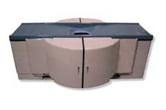
| From left: Image A shows multiplanar and 3D visualization available through cone-beam CT scanning; image B shows a .25-mm sagital cone-beam CT slice with an arrow pointing to a cancer that was not seen on the patient?s mammogram; image C shows patient positioning. Click on breast images for larger versions | ||
The system incorporates a unique horizontal gantry to permit comfortable prone patient positioning and does not require compression of the breast tissue. The breast is suspended through an opening in the table, and hundreds of images are taken in a 360? circle in 10 seconds. Ultrafast image reconstruction allows immediate image review as volumetric images of the entire breast are presented in full 3D and multislice/multiplanar formats. Slice thickness as thin as 0.25 mm eliminates virtually any tissue overlap or superimposition of structures as encountered with 2D projection imaging. Radiation dose is comparable to that of a conventional two-view mammogram and is up to 5 times lower than conventional CT units.
The Spec Sheet
- Spatial and true isotropic resolution: 0.1 to 0.27 mm
- Radiation dose is comparable to a two-view mammogram.
- Scan time: 10 seconds
- Image reconstruction time: less than 2 minutes
- Slice thickness: 0.25 mm
- A horizontal gantry provides prone positioning.
- Breast compression is not required.
The Visible Difference
In an ongoing pilot study at the University of Rochester Medical Center, Koning CT for breast and extremities has demonstrated its potential to equal and, in many cases, outperform current breast imaging methods, according to the company. With high spatial and true isotropic resolution, the system delivers superior image quality and comprehensive diagnostic information as compared to 2D projection x-ray systems.
