|
· A Better Way to X-ray
· Research Alert: Cleveland Clinic Launches Chest X-Ray CAD Study
· Tech Zoom: FCR Go Gives Techs a Helping Hand
A Better Way to X-ray
Biospace Med offers low-dose, high-quality solution
Numerous radiography exams are part of life for the approximately 900,000 people in the United States afflicted with scoliosis. Children, in particular, typically undergo x-ray exams at least twice per year, for 5 to 6 years, in order to monitor the disease’s progression as well as determine treatment effectiveness.
Looking for a solution that would reduce radiation dose without sacrificing image quality, St Justine Hospital for Children in Montreal, Canada, turned to Biospace Med and its EOS ultralow-dose 2D/3D imager.
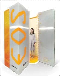 |
| Biospace Med’s EOS has the ability to capture a whole or partial body image in a single scan without any need for digital stitching. |
At the children’s hospital, more than 1,000 pediatric patients with spine deformities, such as scoliosis, have experienced EOS long-length imaging. Biospace Med pointed out that the technology’s use of low dose is especially beneficial for children, who are much more sensitive to the harmful effects of radiation. “EOS can image a patient for 5 years at the dose given to a patient with standard x-ray equipment at their first exam,” explained Richard diMonda, vice president of strategic marketing, global.
Based upon a patented particle-detector technology developed by Nobel Prize winner Georges Charpak, the EOS has the ability to capture a whole or partial body image in a single scan without any need for digital stitching. Biplane images, offering front and lateral views, can be acquired simultaneously in an upright, weight-bearing position.
It also operates on slot-scanning technology, which minimizes the amount of scattered radiation that hits the linear detector. The detector technology, which is based on the charge multiplication effect in pressurized gas, ultimately improves the signal-to-noise ratio and dynamic range. This makes it possible to obtain high-quality images at a lower radiation dose. Furthermore, vertical distortion, commonly associated with x-ray systems, is eliminated, and complete head-to-toe digital images can be produced in 25 seconds or less.
“The low-dose, weight-bearing images offer physicians a global view of the entire body and an understanding of the relationships that exist between joints,” said Jean Dubousset, MD, PhD, a prominent French pediatric orthopedic surgeon who assisted in the design of EOS. “This insight into balance and posture can provide valuable surgical-planning information and reduce the need for higher-dose exams.”
Biospace Med unveiled the EOS to the US market at the RSNA meeting in Chicago. “Anyone who came by the booth to see the system immediately understood its benefits,” diMonda said.
The 2D imager received FDA clearance in September, and Biospace Med plans to apply for clearance of its 3D imager by the first quarter of 2008. Company reps also said they anticipate installations of the EOS imager in the United States by the end of the first quarter.
—Elaine Sanchez
Research Alert: Cleveland Clinic Launches Chest X-Ray CAD Study
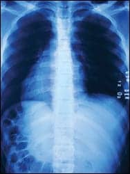
The Cleveland Clinic recently announced the establishment of a 5-year clinical study designed to evaluate the efficacy of chest x-ray CAD at improving practical early detection of lung cancer.
 |
Moulay Meziane, MD, section head of thoracic imaging at The Cleveland Clinic and the principal investigator of the study, explains that the study is important because chest x-ray with CAD could be an economically feasible alternative to screening with CT. “While the usefulness of CT screening is still being debated and investigated, we need to be reminded that CT imaging may not reach or capture as many patients as CR imaging,” Meziane said. “CT scanning is more expensive and will subject the patient to more radiation, and CT machines are not as widely available as CR units.”
But chest x-ray alone has not been shown to be effective for screening, which is where evaluating the efficacy of CAD software comes in. “The results of prior clinical trials that have studied the role of CR for the screening of lung cancer remain unsatisfactory,” Meziane said. “Our purpose is not only to revisit such an issue but also to look at the benefits of CAD and how it may improve the detection of early-stage lung cancer.”
The study will consist of two parts, retrospective and prospective. To evaluate both the performance of CAD for chest x-ray and the performance of clinicians using CAD for chest x-ray, Meziane and colleagues will perform a retrospective study involving three groups of six physicians a piece: general radiologists, pulmonologists, and chest radiologists. “Eighteen physicians with different levels of expertise are testing the CAD systems and we are determining how it affects their interpretation and their clinical management,” said Meziane.
 |
The second component of the study, the prospective portion, will take place over the course of 5 years and will involve 9,000 asymptomatic patients at a high risk for developing lung cancer. “There will be two arms of the prospective study,” explained Meziane. “It’s going to be a blind, randomized clinical trial in which 4500 patients will be subjected to chest x-rays with CAD, while 4500 will not get x-rays or any kind of imaging. They’ll receive what we call a placebo x-ray. They will not know whether they’ve had a chest x-ray or not.”
But even assuming CAD for chest x-ray proves effective in terms of accurately diagnosing early-stage lung cancer, Meziane notes, questions remain about the benefits and risks of using the modality as a mass screening tool. “While we may determine the performance of CAD as a second reader and how it can help the interpreter, we still do not know how it may ultimately affect clinical outcomes and the morbidity and mortality or survival of patients diagnosed with lung cancer,” he said. “Extensive clinical, economical and quality of life studies will be performed on all subjects throughout the trial. Outcome studies will not only look at the individual patients’ results and needs, but also how such results may influence and address public health care at large and local and state concerns.”
The retrospective portion of the study is currently in progress; participants for the clinical trial will be enrolled early this year.
—Cat Vasko
Tech Zoom: FCR Go Gives Techs a Helping Hand
At the 2007 annual meeting of the Radiological Society of North America (RSNA), Fujifilm Medical Systems USA Inc, Stamford, Conn, displayed its new FCR Go portable digital x-ray system. The FCR Go, which is currently awaiting FDA 510(k) clearance, integrates a customized version of Fujifilm’s FCR Carbon XL CR reader and a notebook version of the Flash IIP console with a portable from Hitachi.
 |
“What’s different about FCR Go compared to competitive portable digital systems is its flexibility,” said Penny Maier, national marketing manager with Fujifilm. “You have different sizes of cassettes without a tether cord, and that makes it useful for a number of applications. You can go into the NICU with a smaller-sized cassette, or into the OR with a larger cassette.”
Because its cassettes aren’t hindered by the tether cord required by flat-panel detectors, the FCR Go is lighter and easier to move than traditional portable DR systems. “The weight of the cassette is a big shortcoming with some existing systems,” Maier said. “Fuji cassettes are about half the weight of what a flat-panel detector would weigh. That may not sound like a big deal for one patient, but for technologists who need to image 20 to 25 patients, the weight of cassettes can become pretty demanding physically.”
That’s the real goal of the FCR Go, Maier explained: making life easier for the technologists who operate it. Another useful offering for techs is the “inching” feature. “Inching is a really nice feature to have inside a patient room,” Maier said. “The technologist can make very short movements to enable easier positioning of the patient. The tech can also make adjustments to technique at the tube.”
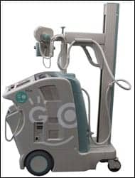 |
| Fujifilm launched its FCR Go portable x-ray system at RSNA. |
Additionally, FCR Go boasts the same functionality and image processing features available at the technologist’s fixed workstation; the portable console offers all the image optimization and processing capabilities of Fujifilm’s Flash IIP.
“This system uses the same imaging plates and cassettes as our other systems,” said Maier, “and it has the same interface and compatibility for the technologist workstation. Just having that consistency really simplifies the process.”
And then there are the advantages everyone in the hospital imaging department can enjoy. “It’s one of the fastest CRs on the market,” noted Maier. “You can see images beginning in as little as 23 seconds. That’s unique against other portable CR devices on the market.
“It has the speed, it has the power, it’s going to be competitively priced,” she continued. “There are few limitations to contend with, which is why it had amazing reception at RSNA. What we’ve learned with hospital-based customers is that there’s a price differential between an analog portable system and a digital portable system, and you can justify the digital as a backup in an emergency if a room goes black. That’s a nice justification for a product like this. It really fits into the whole enterprise. Associated imaging centers and clinics can share in that interface as well.”
Fujifilm anticipates US release of the FCR Go in mid-2008.
—C. Vasko