Ideas in Hospital-Based Imaging
Group Buy Equips Entire City With FFDM
TJU Puts 64-Slice CT in ED
Group Buy Equips Entire City With FFDM
How four competing health care organizations joined forces to improve patient care and drive a better price.
By Dana Hinesly
 |
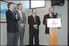
| From left to right are Timothy Charles, president and CEO of Mercy Medical Center; Jerry Rozeboom, MD, of OB-GYN Associates; Ted Townsend, president and CEO of St Luke’s Hospital; Arnold Honick, MD, of RCI at a press conference held at RCI Imaging Center’s patient waiting area. |
In today’s aggressive health care environment, what happened in Cedar Rapids, Iowa, seems almost unthinkable. Four organizations vying for the same patients worked together to focus on the real bottom line: providing the best possible care for their community.
Last September, the area’s four major health care organizations—Mercy Medical Center, OB-GYN Associates, RCI Imaging Center (owned and operated by Radiology Consultants of Iowa PLC [RCI]), and St Luke’s Hospital—culminated a joint effort to acquire and install Selenia digital mammography equipment from Hologic Inc, Bedford, Mass.
“If one hospital were to acquire [new] technology just to get a leg up on another, in very short order, other facilities would obtain the same systems. So, you’re clearly not finding a market share value for a very long period of time by providing that technology,” says Arnold Honick, MD, radiologist at RCI. “By doing it together instead of making it a [competitive] process, we provide a fiscal benefit. We also can put our heads together as far as the technological hurdles that we need to overcome.”
The second largest city in Iowa, metropolitan Cedar Rapids has a population of about 250,000. RCI is the only group practicing radiology for the city, with different components of treatment being provided at all four facilities.
“We technically are competitors; however, all of the facilities use the same radiologists,” says Rebecca Jacobs, MA, RNC, nurse manager of the Women’s Center at Mercy Medical Center. “Because this is a huge work process—not only for the technologists, but in regard to workflow for the radiologists—doing it independently at one site was going to be very complicated.” Also, it is not uncommon for women in Cedar Rapids and surrounding counties to be examined at one facility and receive definitive care at another. “A woman may have her initial screening done at OB-GYN Associates, her diagnostic workup completed at RCI, and then the surgery performed at Mercy Hospital,” Jacobs explains.
Making the Choice
A decision-making committee was formed in October 2004, primarily as an informational venture, providing an opportunity for Honick to convince the hospital that digital mammography was going to be beneficial to the community as well as benefit both radiologists and providers. The committee was made up of administrators, managers, technologists, and IT staff from each of the four sites, all of whom were involved in every aspect of the project.
“The meetings [then became] more evaluative, and they involved lots of input,” says Kathryn Epley, chief administrative officer at RCI. “We went on site visits, then selection, then implementation. It became a natural progression of planning and working together without any site gaining an advantage, with the focus of improving women’s health in the community.”
Initially, there was some resistance—both to the necessity of digital mammography as well as to the collaboration with competing hospitals—much of which could be attributed to the lack of proof that digital imaging provided improved cancer detection capabilities as compared to film screen.
That all changed when the results of the Digital Mammographic Imaging Screening Trial (DMIST)—a 4-year study sponsored by the National Cancer Institute—were published. The research indicated that in women younger than 50, digital mammography detected about 15% to 28% more cancers than with traditional, film-based mammography. Improved results also were found in women with dense breasts and in premenopausal women, for whom digital capture detected 15% more and 21% more cancers, respectively.
“Working together allowed us to put all of our heads together, putting the best of the best to work and enabling us to come up with answers that always kept the customer foremost in our minds,” says Kristi Thomson, RT(R)(M)(QM), CDT, BSHCA, supervisor of the Women’s Breast & Bone Health service at St Luke’s Hospital. “I believe that more and more organizations will collaborate in the future, because the benefits far outweigh any negatives.”
Money Talks
During the 2 years of meetings and discussions, each of the organizations earmarked funding to make the capital investments necessary when the time came. Working together also proved a proficient way of paying less for each gantry.
“Individually, each facility received quotes from vendors, but the respective buying groups were unable to beat the price that we accomplished through negotiations by purchasing the units simultaneously,” says Honick, who estimates a 10% to 15% cost savings on each gantry.
The total capital investment for all seven systems was more than $3 million. “The other benefit we received was the accessories you need to have, such as the workstation, the CAD, and the things that go with making it a complete package,” he notes. “There were substantial savings on everything, including [those types of] accessories.”
Making Progress
The transition has not been flawless; some bugs are being worked out that are slowing the transfer of images.
“The rapid and total implementation of digital mammography was difficult and stressful to all involved. It has been perhaps most stressful for the radiologists in learning an entirely new workflow and performing critical interpretations using a combination of film and digital images,” says Epley, who adds that, nevertheless, the new digital breast images are stunning in their clarity and that the clinicians appreciate the computer tools available to aid radiologists with interpretations. “The trade-off is that … managing the studies in both film and digital form also has added greatly to the amount of time clerical staff is required to devote to preparing the studies to be interpreted. We do expect this to improve over time.”
Dana Hinesly is a contributing writer for Axis Imaging News. For more information, contact .
TJU Puts 64-Slice CT in ED
Patients with chest pain and trauma victims benefit from speeded throughput and a reduction in unnecessary hospitalizations
By Renee Diiulio
 |
| Vijay Rao, MD, FACR, Thomas Jefferson University Hosptal |
Place a CT scanner in the emergency department (ED), and physicians will use it. Research has shown that CT utilization increases when the equipment is available in the ED. In fact, a paper presented at RSNA 2006 reported that growth in the utilization rate of CT in the ED outpaced that of patient volume.1 The researchers found that from 2000 to 2005, adult ED patient volume increased by 13% while head CT increased by 51%, cervical spine CT by 463%, chest CT by 226%, abdominal CT by 72%, and miscellaneous CT by 132%.1
Although the team did not collect data on the resulting impact on patient volumes, another group did. Beland et al examined the volume of trauma CT and diagnostic yield in a busy level I trauma ED over 6 years.2 The researchers found that the volume of examinations rose by 6% and diagnostic yield by 3%. Their conclusion: “This study demonstrates a concomitant increase in the diagnostic yield for these examinations.”2
These presentations had not been made when Thomas Jefferson University Hospital (TJU), Philadelphia, installed a Brilliance 64-slice CT from Philips Medical Systems, Andover, Mass, in the ED in October 2006. But by December, the hospital had already seen immediate improvements. “We have improved patient care, convenience, and throughput,” says Vijay Rao, MD, FACR, professor of radiology and department chair at TJU. She attributes a much shorter turnaround to the associated reduction in patient transport, and a decrease in unnecessary hospitalizations to the faster and more accurate diagnosis.
A review of the literature by Matthew Budoff, MD, associate professor of medicine in the division of cardiology at the Los Angeles Biomedical Research Institute of Harbor–University of California, Los Angeles, Medical Center, Torrance, Calif, found that “multi-slice CT has the potential to more effectively triage patients who present to the ED with chest pain.”3 Rao notes that it was the desire to provide cardiac CT service to patients presenting with chest pain that led the department to select a 64-slice CT. “We wanted to perform CTA examinations on low- and intermediate-risk patients to prevent unnecessary admissions and reduce patient waiting time,” she says.
Patients whose coronary CTA results are completely negative are discharged. “They do not undergo serial enzymes, they don’t wait 6 hours, and they avoid unnecessary hospitalization,” Rao explains.
Although the major driving factor was cardiac, trauma patients have benefited from 64-slice CT as well. “It’s wonderful to have a fast scanner for level I trauma patients,” Rao says. “We can scan large fields very quickly.”
The workflow process remains similar. ED physicians supervise the contrast injection, but the radiology department continues to manage the scanner, throughput, protocol, and readings. “The ED physicians are using the service no differently than when the exams were performed in the main radiology department,” she says. Patients, however, are no longer transported from the ED on the first floor of one building to radiology on the third floor of a connected building.
“Because we were fairly removed from the ED, there was a lot of time wasted transporting patients. And it required additional resources, because those who are critically ill require supervision,” Rao says. The ED had always expressed interest in having a CT scanner in the department, but the decision was left to radiology.
Rao felt that the new CT would allow the department to expand its capacity, which was needed, and improve its service to the ED. Because the service is so new, Rao has not yet collected data that illustrates the benefits, nor has she gathered utilization data. “We hear that a CT in the ED causes utilization to skyrocket, but we have not seen a substantial increase yet,” Rao says.
To keep the number of examinations reasonable, the department is informally educating ED physicians on the radiation risks of CT, but Rao would like to see more formal training take place throughout the enterprise. “It’s our role to educate the ED physician about radiation risk so that CT is not overutilized,” she says, noting that the hospital is currently examining workflow processes within the institution for additional process improvement.
“We are one piece of the entire pathway that the patient has to go through, so it’s a team approach. We’re trying to fine-tune our processes to reduce length of stay in the ED,” Rao notes. By eliminating such steps as transportation, they have taken a big step in improving patient care and workflow.
Renee DiIulio is a contributing writer for Axis Imaging News. For more information, contact .
References
- Broder J, Warshauer D. Emergency radiology (imaging applications in trauma and emergency care II): increasing emergency department utilization of computed tomography: 2000-2005. Session presented at: Annual Meeting of the Radiological Society of North America; November 27, 2006; Chicago. Available at: rsna2006.rsna.org/rsna2006/V2006/conference/event_display.cfm?id=66601&em_id=4427881. Accessed on January 12, 2007.
- Bleland M, Mayo-Smith W, Egglin T, Murphy B, Golding D, Biffl W. Emergency radiology (imaging applications in trauma and emergency care II): CT utilization in a level I trauma center over 6 years: have indications and diagnostic yield changed? Session presented at: Annual Meeting of the Radiological Society of North America; November 27, 2006; Chicago. Available at: rsna2006.rsna.org/rsna2006/V2006/conference/event_display.cfm?id=66601&em_id=4435537. Accessed on January 12, 2007.
- Budoff M. Cardiac CT in the emergency room. Applied Radiology. 2006;35(suppl)(12):48–55.


