 |
Ideas in Hospital-Based Imaging |
Molecular Approach in Breast Diagnostics Shows Promise
Johns Hopkins Installs First 320-Slice CT in the United States
Molecular Approach in Breast Diagnostics Shows Promise
Nearly 3 years ago at the National Consortium for Breast Centers meeting in Las Vegas, vendors opened Leora Lanzkowsky’s eyes to Breast-Specific Gamma Imaging (BSGI), a novel molecular imaging approach in breast diagnostics that employs a high-resolution small field-of-view gamma camera and a radiotracer.
 |
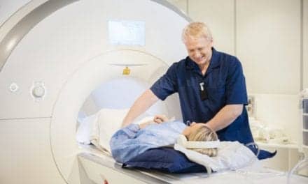
| The portable Dilon 6800 Gamma Camera provides the ability to image up to the chest wall and mimic all mammographic views. |
Since that first introduction, the medical director of the Eisenhower Schnitzer/Novack Breast Center in Rancho Mirage, Calif, has seen the technique’s emergence as a solution for many difficult-to-diagnose patients. “I can see a huge future for this technology,” Lanzkowsky said.
Under BSGI, radioisotope Tc-Sestamibi is taken up by cells with increased metabolic activity, such as rapidly dividing breast cancer cells. Highly focalized areas of radiotracer uptake indicate areas of concern called hot spots. This process replaces the more limited previous method employed known as scintimammography.
“The need for the technique came about on several fronts,” Lanzkowsky said. “For example, the realization that high-risk women need additional adjunctive imaging and that mammography has limitations, especially in women with dense breast tissue. The addition of a functional study to a morphologic study increases the pretest predictability in a very useable way.”
Furthermore, Lanzkowsky said, the procedure is helpful in women with severe fibrocystic changes and multiple masses detected by ultrasound. Eliminating “background noise,” BSGI is sometimes ordered by the MRI reader to decrease false positives. It is also useful in women who are 85 years or older, suffer from claustrophobia, or have pacemakers or renal function issues. BSGI can avoid unnecessary biopsies in this clinically fragile group of women, Lanzkowsky said.
If a suspicious finding on mammography or ultrasound is discovered, and the BSGI is negative, Lanzkowsky said she would schedule a 6-month follow-up with the patient instead of biopsy. Likewise, if there is extreme density or findings that would normally lead women to additional mammography and ultrasound, a BSGI is performed immediately after the screener. If results come back negative, an appointment is scheduled for the next year.
Lanzkowsky said her patients have embraced the technology. The procedure is both comfortable and easy for patients to understand. “The patients are shown all their imaging, and it is very easy for them to comprehend the results of the BSGI scan,” she said.
According to Nancy F. Morter, director of marketing and corporate communications at Dilon Technologies, BSGI provides not only physical benefits, but also financial rewards.
 |
| High-risk patients with dense breast tissue are benefiting from BSGI. |
“The economic impact can be significant for hospitals and insurance companies because as a diagnostic breast imaging test, BSGI has comparable sensitivity to and greater specificity than MRI, which for clinics equate to fewer false positives,” said Morter, whose company manufactures the small, portable Dilon 6800 Gamma Camera with the ability to image up to the chest wall and mimic all mammographic views. “BSGI has an extremely high NPV and costs significantly less than an MRI.”
At last November’s RSNA meeting, study results were presented demonstrating high predictive values of BSGI for identifying breast cancer. Led by Jean Weigert, MD, director of breast imaging at Mandell and Blau MDs PC in New Britain, Conn, the study reviewed 512 women referred to BSGI after an indeterminate mammogram or ultrasound or a history of breast cancer; 81% of the patients had 6- to 24-month follow-up with no new findings. Additionally, 97 biopsies were performed, of which 46 were positive and 51 were negative, five in patients with negative studies.
Lanzkowsky also looks forward to publishing her own findings, which point to BSGI as “a reliable and well-tolerated shortcut to the detection of significant lesions.”
BSGI is currently performed in many leading medical centers around the country, including Cornell University Medical Center in New York, George Washington University Medical Center in Washington, Northwestern Memorial Hospital in Chicago, and The Rose in Houston.
—Elaine Sanchez
Johns Hopkins Installs First 320-Slice CT in the United States
The Johns Hopkins Hospital, Baltimore, recently became the first medical institution in the United States to install the Aquilion ONE 320-slice CT scanner from Toshiba America Medical Systems Inc, Tustin, Calif. The 2-ton device, which costs more than $1 million, is one of two devices in the United States to be installed this year and is currently operational. It was awarded FDA clearance on November 27, 2007.
 |
| The Aquilion ONE’s detector coverage is more than five times greater than that of a 64-slice scanner. |
The 320-slice scanner’s detector coverage is more than five times greater than that of a 64-slice device, and it can capture slices as wide as 16 cm in less than 1 second. “Full cardiac coverage with 320 detectors allows for the lowering of the radiation dose and improved temporal resolution, but there are several other advantages,” explained Richard George, MD, assistant professor of medicine in Johns Hopkins’ cardiology division. “The ability to image the heart over one heartbeat or less eliminates the need to reconstruct images over multiple heartbeats into a full cardiac volume. This eliminates step artifacts often seen with other scanners. Shorter scan times require lower contrast doses and fewer opportunities for motion artifacts secondary to patient movement.”
George notes that the 320-slice device shows promise for multiple cardiac applications, including the imaging of arrhythmias and stress perfusion imaging. “Full cardiac coverage over one heartbeat allows for imaging of atrial fibrillation and premature beats,” said George. “Furthermore, the lower contrast and radiation dose opens up the potential for rest and stress perfusion imaging, a promising application using cardiac CT.”
The development of 320-slice CT has been a long time coming; the Aquilion ONE is currently the only scanner on the market to boast such a wide detector area. The system utilizes the industry’s thinnest detector elements, just 0.5 mm, and is equipped with a table that accommodates patients weighing up to 650 pounds and coneXact dynamic volume CT reconstruction. Prototype 256-slice scanners were tested both in Japan and at Johns Hopkins prior to the release of the 320-slice device.
“The 320 system is a striking improvement over the 256 prototype systems,” said George. “While 256 detectors will cover most patients’ hearts, the extension of the detector array from 256 to 320 detectors ensures that every patient will have their entire heart covered by the detector array. We’re also very impressed with the improvements in processing time.” George says the team at Johns Hopkins have also significantly reduced dosage, thanks to the scanner’s improved resolution and imaging time: most CT angiograms require less than 5 mSv, depending on heart rate.
According to George, it’s still too early to make a call regarding the economic efficiency of such an expensive, high-powered device. Because it can safely image the whole brain, there are exciting implications for imaging of patients exhibiting stroke symptoms. The Aquilion ONE can rapidly acquire complete functional data with less radiation and contrast, speeding diagnosis. And patients with atrial fibrillation or frequent premature ventricular beats can now have a noninvasive angiogram using the system. “It is expected that the 320-slice CT scanner will be more accurate and applicable to a more diverse and complicated patient population than 64-detector technology,” said George.
—Cat Vasko