For many decades, the X-ray angiogram has been the gold standard in evaluating the patency and function of veins and arteries, the effectiveness of surgical and medical treatment as well as diagnosing vascular disease. Yet now, CT, MR and electron beam angiography are offering powerful alternatives to ?ole reliable X-ray angiography.
Today, angiographic studies utilize the entire range of radiographic modalities including X-ray, magnetic resonance (MR), computed tomography (CT) and electron beam CT scanners to diagnose conditions that reduce the flow of blood through arteries that supply the heart, kidney, brain or peripheral area of the body.
While each imaging technique presents advantages and disadvantages, decisions about which method to employ hinge on issues such as the clarity of images produced for diagnostic purposes balanced against the level of invasiveness that the patient experiences. New capabilities in producing three-dimensional images upgrade diagnostic and treatment options. While the costs associated with these techniques are certainly a critically important consideration, the overriding concern seems to be improved management of disease using the least invasive methods. Emerging trends suggest that we’ll see increased use of MRA and CTA or in some instances electron beam scanning for diagnosis and interventional planning, while reserving conventional angiography for interventional procedures.
Comparing Techniques
“The highest spatial resolution is obtained with X-ray catheter or ?conventional’ angiography, and this represents the reference standard to which the other techniques are compared,” explains Mark Delano, M.D., assistant professor in the Department of Radiology at Michigan State University. “With greater spatial resolution, there is a greater ability to discriminate or identify smaller structures. This is of critical importance in the evaluation of intracranial aneurysms or narrowing of blood vessels.”
Conventional X-ray angiography offers the advantage of facilitating both diagnosis and intervention in one procedure. Geoffrey Rubin, M.D., assistant professor of radiology at Stanford University School of Medicine, notes, “When using conventional angiography, you are right there and can do something if it is necessary as you see the abnormality. You can assess pressure gradients in real time.”
 But in 2000, variations on the conventional are adding new dimensions, literally. Three-dimensional rotational angiography, such as that done on the Integris system (made by Philips Medical Systems North America, Shelton, Conn.) uses a conventional C-arm with enhanced hardware and software to magnify structures being studied and create them in 3D, explains John Steidley, director of the Vascular Business Unit at Philips. Integris is capable of spinning 180 degrees around the patient while acquiring data throughout the rotation. Once the images are obtained, the system uses a CT reconstruction algorithm to complete the three dimensional picture of contrast filled blood vessels. It can be used in any part of the body that can be held motionless for eight seconds, such as the head.
Controlling the Invasion
The primary disadvantage to conventional angiography is the level of “invasiveness” required, Michigan State’s Delano says. In traditional studies, a catheter is inserted into a major artery (usually the femoral) and is advanced to the section of the body to be studied, where a contrast agent is injected to perform the examination. “The potential for damage to the patient’s arteries, stroke or other embolic complication from the procedure is real and depends on the region of the body to be evaluated,” Delano continues. “For example, the risk of stroke from catheter angiography for evaluation of the carotid arteries is estimated between 1 in 1,000 and 1 in 3,000 depending on the age of the patient and the skill of the angiographer, among other factors. The actual complication rate may be small, but the consequences are devastating, if not catastrophic, for the unlucky individual involved.”
One means of reducing the complication rate involves the use of alternative imaging techniques, such as CTA and MRA. “We use CTA between five and eight times per day in our department to image the aorta and its branches, patients with aneurysms, stent graphs, renal artery stenosis and carotid occlusive disease,” says Stanford’s Rubin. Because the contrast medium is administered via peripheral intravenous route, there is no need to insert an arterial catheter, thereby decreasing the complications inherent in arterial catheter placement.
In addition, Rubin notes that three-dimensional capabilities of CTA offer an increased diagnostic capacity in the management of aneurysms. “We can decide what size graft to use prior to surgery, based on the imaging,” he says.
Using volumetric data provided by CTA, the surgeon is able to plan better for the surgical graft to use for repairing such defects as an aneurysm. Rubin says that in interventional planning, CTA offers increased clarity to differentiate tissue types. For example, in a traditional angiographic examination of an aneurysm, it may be difficult to distinguish soft plaque inside the vessel from the outer wall of the vessel. CTA provides enhanced visualization capability.
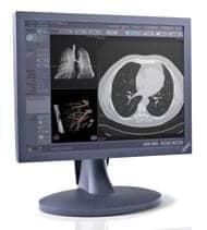
 This CT angiography image was created on a GE LightSpeed scanner
Russell Golkow, M.D., clinical director of CT at the Kennedy Health Systems, in Cherry Hill, N.J., is another proponent of CTA. Since installation of their GE Medical Systems (Waukesha. Wis.) LightSpeed CT scanner in September 1999, they have essentially replaced their conventional diagnostic angiography program with CT angiography.
“We are finding we’re getting better images than we were with our old angiography [method]. With an IV injection in the patient’s vein, we can do a 30-second CT scan at one tenth the cost of traditional angiography,” Golkow says.
He indicates that CT scans are now used as a diagnostic tool to improve decisions about which course of action should be taken, and to plan the exact area of intervention required. “For those patients who need intervention, the CTA does a diagnostic work-up so we can go to the area of disease in less time with decreased risk of complications,” he says.
So has CTA reduced Kennedy Health Systems’ use of traditional angiography? Not really. “We have found this has no negative impact on the number of traditional angiograms we do,” Golkow replies. “We use CTA as a portal exam, and believe it will increase the number of patients scanned.” In fact, he notes that the facility has almost doubled its volume in two months.
Golkow remarks that Kennedy has experienced a steep learning curve for technicians due to the complexity of the equipment. He and his staff have developed a series of protocols to assist in interpreting findings and developing techniques. “Because the scanner is so powerful at collecting data, we are now doing things we never did before, like angiography,” he says.
The MR Method
Just like CTA, MRA too is gaining believers. “MRA is a fantastic screening examination and is rapidly becoming the definitive evaluation in many forms of vascular disease,” Delano asserts. “This role is expanding as better equipment and techniques are becoming available in the community and as physicians become more aware of the available techniques.
“Recent improvements in MRA techniques are allowing for much greater resolution than available previously and image quality is now occasionally mistaken for conventional angiography,” Delano continues.
 This MRA image of a male patient with a
suspected aneurysm and thrombus in the lower left femoral artery utilizes Epix Medical’s AngioMARK imaging agent.
One of the advantages provided by both CTA and MRA is their 3D capability that permits images to be manipulated so that the physician can visualize all planes of the anatomical structure being studied. At Beth Israel Medical Center in New York City new endovascular techniques are being carried out with a new 3D Virtuoso computer program developed by Siemens Medical Systems (Iselin, N.J.).
“We use this system for endovascular techniques, because the stereoscopy gives us the ability to see depth,” says Alejandro Berenstein, M.D., director of the Institute for Neurology and Neurosurgery at Beth Israel. The system uses a special monitor combined with polarized glasses worn
by the operator that provides stereoscopic views of reconstructed images.
“Siemens is the only manufacturer with this stereoscopic reviewing capability,” Berenstein adds. There are presently only two sites in the United States working with 3D Virtuoso. In his center, Berenstein is using 3D Virtuoso to treat cerebrovascular disorders and vascular conditions of the head, neck, brain and spinal cord.
Electron Beam Tomography
Yet another method of angiography, electron beam angiography, is gaining popularity too. This application is intended to function as a diagnostic X-ray system that can produce two- and three-dimensional images of the heart, blood vessels and lymphatic system.
 Imatron’s C-150 EBT scanner illustrates a heart with three bypass grafts.
“We are confident that coronary EBA, which, because of our scanner’s speed and ability to ?freeze’ heart motion is unique to EBT,” explains S. Lewis Meyer, CEO of Imatron, Inc. (South San Francisco, Calif.), the manufacturer of the Electron Beam Tomography (EBT) scanner.
“In EBA, the scan is perfectly registered and synchronized to the EKG which means all images are produced in the same phase of a heartbeat,” Meyer says. “We can actually visualize the lumen of a coronary artery in a procedure that takes 20 minutes to complete.”
One of the many potential uses for EBA is in the evaluation of the function of coronary bypass grafts and of other patients who would not ordinarily have follow-up studies to evaluate the effectiveness of treatment.
“Our typical patient is a person with a previous stent or angioplasty with an abnormal stress test, or abnormal EKG without symptoms, but where there is a question of whether or not he or she has had a heart attack,” explains Matthew Budoff, M.D., director of the Electron Beam Laboratory at Harbor-UCLA Medical Center and assistant professor of medicine at UCLA. “Already our cardiologists are using EBA in place of traditional angiography.”
Budoff notes that with EBA, the patient receives less radiation, and contrast media is injected into a peripheral IV. “Since a number of our patients have had an angiogram before, they therefore know they are not allergic to the contrast agent,” he concludes.
As an imaging technique, EBA is three to five times faster than traditional CTA, according to Budoff. “Traditional CT will get motion artifacts: the coronary arteries look blurred as the heart moves,” he says. “Besides the speed of the actual imaging examination, the entire test takes 30 minutes. In three hours, we can do four to six cases,” Budoff says.
William Stanford, M.D., professor of radiology in the Department of Medicine at the University of Iowa, has been involved in research investigations of the use of the triggered EBT scanner. He notes new developments offer exciting potential uses for MRA, CTA and EBA, but cautions that the collection of more data is necessary for evaluation of the appropriate use of all of this technology. “While there are many exciting developments being pursued,” Stanford concludes, “further studies are necessary.” He raises ethical concerns about determining the best imaging techniques to be used for each patient.
Another dilemma physicians must confront when deciding which imaging modality to employ involves the use of contrast agents to intensify images. Conventional angiography, EBA and CTA use an iodinated contrast agent that can be toxic to the kidneys. Therefore any patient whose renal function is compromised would be a less viable
candidate for using these angiographic procedures. MRA contrast media has no renal toxicity. However, given the use of huge magnets in MRA, patients
with pacemakers, defibrillators or metal implants usually are unable to have these scans performed.
Cost Considerations
No discussion of angiography would be complete without considering the economic implications of the various imaging techniques. When comparing relative costs between imaging techniques used in angiography, a primary consideration returns to the issue of invasiveness. Because a conventional angiogram requires catheter placement into a major artery, the patient requires careful monitoring both during and following the procedure for a number of hours to reduce opportunities for life-threatening complications. Inpatient management is automatically more expensive than tests that can be performed in an outpatient setting.
One of the factors in cost comparison between techniques involves the staffing requirements. In conventional angiography, a patient procedure is usually staffed with a physician, a nurse and a technician who are all required to spend several hours completing the study and evaluating the patient’s condition post procedure. In their department, using CTA their staffing requirements include one physician and one technologist. The test usually takes less than 30 minutes of staff time once the technician and radiologist have learned the techniques.
Imaging studies are usually covered in most insurance plans. Michigan State’s Delano explains that “both CTA and MRA cost on the order of $500 to $1,000, while a conventional angiogram costs several thousand dollars, depending on the area imaged.” Harbor-UCLA’s Budoff says that his department has used the Imatron EBT scanner to complete about 1,000 studies. “We charge approximately $1,000 for EBA. Conventional angiography can run between $3,000 and $6,000, depending on where the study is done.” Golkow of Kennedy Health Systems says that utilizing CTA, “our 30 second scan is about one-tenth the cost of traditional angiography and between one third and one half the cost of MRA.”
Advances in technologic capabilities across the spectrum of imaging equipment continues to improve the clinicians’ ability to diagnose and treat vascular disease. The use of three-dimensional images, ever-faster data acquisition and analysis, and enhanced capability to review an entire patient’s vascular situation provides exciting possibilities to improve patient care. ![]()