Researchers from the WVU Cancer Institute develop new technology for head and neck cancer imaging
Raymond Raylman, vice chair of research for the Department of Radiology at West Virginia University and a member of the WVU Cancer Institute, developed a scanner that combines positron emission tomography and X-ray computed tomography for head and neck cancer imaging.
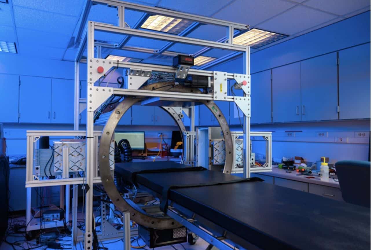
“What makes this system unique is that it has much higher resolution than the standard PET/CT system,” explains Raylman. “That’s because it’s designed specifically for head and neck cancer imaging. The typical scanner systems are general. They’re designed to image brain tumors, breast tumors, and prostate cancer. Instead of doing that, we decided to design one just for head and neck cancer.”
According to Raylman, the reason head and neck cancers tend to be so hard to treat is their size. By the time the typical patient receives a diagnosis, “the tumor can often be centimeters in diameter.”
“This system is designed for estimating the edges of the tumor as accurately as possible,” Raylman shared. “The more accurately you can assess the size, shape and spread of the tumor, the more accurately—and hopefully, effectively—you can plan either surgery or radiation therapy.”
He and his team recently tested their system by using it to produce highly detailed images of simulated head and neck cancers. They are now reportedly seeking regulatory approval to test its performance on 40 cancer patients.
The prototype was funded by the National Cancer Institute, which awarded the project $1.9 million over the course of five years.
The project is a collaboration between Raylman and his colleagues in the School of Medicine, advanced manufacturing experts from the Benjamin M Statler College of Engineering and Mineral Resources’ Lane Innovation Hub, and Xoran Technologies, a manufacturer of X-ray CT scanners.
“This is exactly the type of collaboration that the Hub was designed for,” commented Kelsey Crawford, shop manager for the Innovation Hub’s Advanced Manufacturing Lab. “Having these capabilities on campus allows researchers from all disciplines to create and iterate their designs, and that’s what we did with this technology.”
According to research provided by the CDC, data from 2012 to 2016 shows that approximately 44,000 Americans are diagnosed with an HPV-associated cancer every year. About 19,100 of those people are men, and the cancers they’re diagnosed with most frequently are in the head and neck.
Reportedly, one of the biggest risk factors for developing cancer of the head or neck is contracting the human papillomavirus, a very common viral infection that’s transmitted sexually.
According to the Centers for Disease Control and Prevention, nearly everyone will get HPV at some point in their lives. Nine times out of 10, the virus goes away without causing any problems. But in the other 10% of cases, the virus persists and can cause long-term health issues, including cancer.
“HPV-associated cancers in the oropharynx are on the rise, and many of those spread to lymph nodes before they are large enough to be detected,” explained The Cancer Institute’s Tanya Fancy, chief of head and neck surgery at WVU Medicine.
“A more sensitive, higher-resolution PET scan like this one can help identify the source of the cancer and allow for a more focused treatment. It would define tumor borders and tumor volume more precisely and show the presence of cancer in lymph nodes in the neck more accurately. Both aspects are crucial to identifying the stage of a patient’s cancer and planning for surgery,” Fancy concluded.