Experts study the usage of machine learning and MRI for prostate cancer diagnosis and prognosis
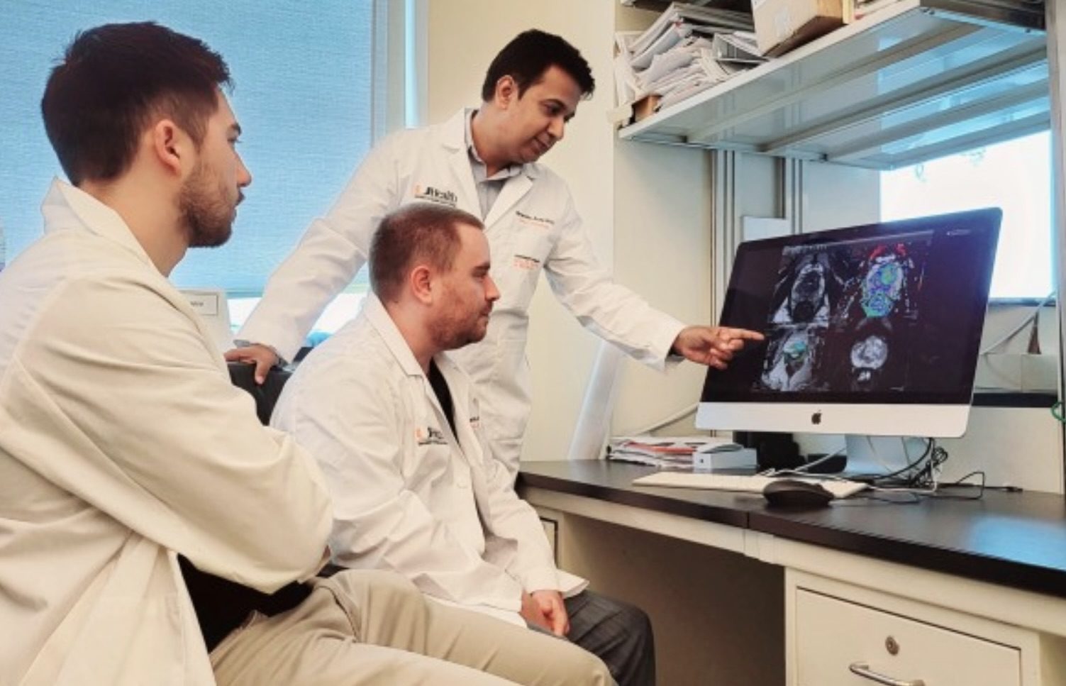
Researchers at Sylvester Comprehensive Cancer Center and the Desai Sethi Urology Institute are studying machine learning for the diagnosis and prognosis of prostate cancer using MRI.
The recent integration of open-source data with machine learning models, especially in the medical field, has opened new doors to studying disease progression and/or regression, according to experts. The use of conventional MRI in prostate cancer research and treatment is effective for prognosis, diagnosis, active surveillance, and reducing the need for biopsy procedures in lower-risk patients.
“Recent advances in machine learning suggest there is promise in developing pipelines that could automate standardized and objective assessments from MRI images while reducing time, human capital, and other resource costs,” explained Himanshu Arora, PhD, assistant professor at Sylvester and the Desai Sethi Urology Institute at the Miller School of Medicine.
According to experts, there are obstacles to using machine learning in patient care, including using machine learning approaches for specific cancers, the specificity of training data for a particular medical condition, etc. In this context, the most recent technologies, such as generative adversarial networks (GANs), are being studied as a potential way to generate synthetic data that preserve the clinical variability of a condition and be applied to PET, CT, MRI, ultrasound, and x-ray imaging in the brain, abdomen, and chest. However, the GAN model usage reportedly remains minimal when depicting the heterogeneity of a disease such as prostate cancer.
“The translational research team is focused on improvising the GAN tools to allow the output images to be applied and integrated with diagnostic and prognostic tools in prostate cancer, “ Arora shared. “Timely diagnosis and assessment of prognosis are challenges for prostate cancer, and this results in many deaths and increases [risk of disease progression],” Arora explained. “We cannot replace the human eye when it comes to medical decision-making. Still, the improvement in technologies could potentially assist radiation oncologists in making timely decisions.”
GAN reportedly provides a long-term impact on evolving the machine learning models by requiring less data and patient follow-ups for effective predictions. Researchers share that the rationale for using GAN is to use machine learning capabilities and generate digital images by learning from previous follow-ups (MRI images and clinical parameters) and understanding and predicting disease progression or regression patterns.
“Technically, the technology developed here is the first start to building more sophisticated models of ‘data augmentation’ where new digital images can be used in further analysis. This is an early phase of our study, but the outcomes are extremely promising,” Arora concluded.