Summary: A new hand-held scanner developed by British researchers can generate detailed 3D photoacoustic images in seconds, enabling real-time diagnosis of diseases like cancer and arthritis.
Key Takeaways
- The new hand-held scanner developed by UCL researchers generates 3D photoacoustic images in seconds, potentially allowing for earlier disease diagnosis.
- This technology, which uses laser-generated ultrasound waves, significantly improves imaging speed and quality compared to previous photoacoustic tomography (PAT) systems.
- Pre-clinical tests demonstrated the scanner’s ability to provide detailed images of blood vessels, aiding in the early diagnosis and monitoring of conditions like diabetes and cancer.
—————————————————————————————————————————————————————————————
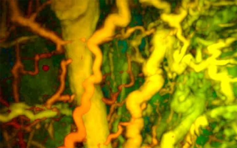
A new hand-held scanner developed by University College London (UCL) researchers generates detailed 3D photoacoustic images in seconds, potentially enabling earlier disease diagnosis in clinical settings. The study, published in Nature Biomedical Engineering, demonstrates that this technology delivers photoacoustic tomography (PAT) imaging scans in real time, offering intricate images of blood vessels.
Photoacoustic tomography uses laser-generated ultrasound waves to visualize changes in veins and arteries up to 15mm deep. Previously, PAT technology was too slow to provide high-quality 3D images, requiring patients to remain motionless for up to five minutes. The new scanner reduces imaging time to a few seconds, significantly improving image quality and suitability for frail patients.
Photoacoustic Imaging
“The breakthrough in this study is the acceleration in the time it takes to acquire images, which is between 100 and 1,000 times faster than previous scanners,” says Paul Beard, PhD, a professor in medical physics and biomedical engineering at UCL. “These technical advances make the system suitable for clinical use for the first time.”
In pre-clinical tests, the scanner produced detailed 3D images of microvasculature in the feet of patients with type-2 diabetes, highlighting vessel deformities. It also visualized skin inflammation linked to breast cancer.
Andrew Plumb, MD, an associate professor of medical imaging at UCL, adds: “Photoacoustic imaging could give us much more detailed information to facilitate early diagnosis, as well as better understand disease progression more generally.”