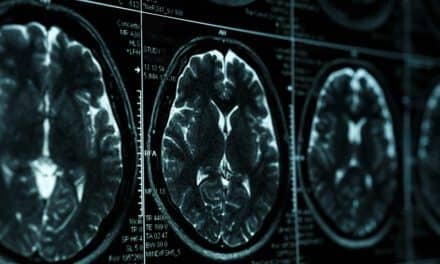
In a case published in the Journal of Intensive Care Medicine, a 54-year-old man was admitted to a hospital in Marseilles, France, with suspected SARS-CoV-2 infection. A nasopharyngeal sample was obtained for PCR analysis, and a low-dose thoracic CT scan and a lung ultrasound were performed within a short amount of time. The PCR showed that the patient was positive for SARS-CoV-2 infection.
A direct comparison of the contemporaneous CT and ultrasound scans show similar findings regarding the locations and patterns of the disease within the lungs. The authors note, “Lung ultrasonography may be considered a useful alternative to low-dose chest CT for diagnosis and management of COVID-19 given its ease of use, repeatability, reproducibility, absence of radiation, and immediate bedside application that obviates the need to transport the critically ill patient to the CT scanner.”
Find the paper in the Journal of Intensive Care Medicine.
Featured image: The transverse thoracic CT scan image shows multi-lobar asymmetric lung lesions with peripheral distribution of ground glass opacities, consolidation, and crazy pavement pattern. The lung ultrasonography is presented as thumbnail images that correspond to different areas of the CT scan indicated with long yellow arrows. A and B show A lines (normal aeration pattern); C and D show focal and confluent B lines (interstitial pattern); E and F show thickening and irregularity of the pleural line in association with B lines (suggesting primary lung injury as the cause for the B lines). B lines and pleural irregularity are indicated with short yellow arrows. Courtesy, Journal of Intensive Care Medicine