Steven C. Horii, MD (center) is flanked by two PACS pioneers who worked on early digital radiography and teleradiology projects at UPenn: (left) Dan Morton, manager of medical informatics, UPenn, and Jack W. London, PhD, (right) director, Kimmel Cancer Center Shared Computer Facility at Thomas Jefferson University, Philadelphia. Steven C. Horii, MD (center) is flanked by two PACS pioneers who worked on early digital radiography and teleradiology projects at UPenn: (left) Dan Morton, manager of medical informatics, UPenn, and Jack W. London, PhD, (right) director, Kimmel Cancer Center Shared Computer Facility at Thomas Jefferson University, Philadelphia. |
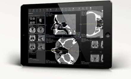
Any image, anytime, anywhere—that’s the mantra,” says Reuben Mezrich, MD, PhD, describing the capability of the modern PACS (picture archiving and communications system). “But none of this could have happened without DICOM (digital imaging and communication in medicine).
“If you could give a Nobel Prize for DICOM, that would be a good thing,” adds Mezrich, professor of radiology and chairman of the radiology department at the University of Maryland School of Medicine, Baltimore.
DICOM is a meticulously developed set of standards that allow systems to interface. It specifies how devices built in conformance with the standards react to commands and data being exchanged. DICOM, for instance, lets a CT scanner made by one manufacturer, an MRI scanner made by a second company, and an ultrasound machine made by a third company all communicate with the same PACS. It is because of DICOM that images from all three modalities, and others as well, can be displayed and interpreted at the same PACS workstation. The images can all be sent to the same PACS archive. DICOM is the computer standard that lets a PACS do its work.
If, as Mezrich suggests, a prize were given for DICOM, the recipient would most likely be Steven C. Horii, MD, who is now a professor of radiology and clinical director of medical informatics at the Hospital of the University of Pennsylvania, Philadelphia. By informal acclaim from his peers, Horii is credited with being the DICOM point man. He is cited for putting in the long hours and the blood, sweat, and perseverance that were necessary to DICOM’s creation.
“Horii was one of the really fine leaders in DICOM. He is very important in the development of DICOM,” says Mezrich.
 Andre Duerinckx, MD, PhD Andre Duerinckx, MD, PhD |
Says Andre Duerinckx (pronounced Dur-inks), MD, PhD, himself a PACS pioneer and now director of cardiac MR and CT and nuclear medicine at Forsyth Radiological Associates in Winston-Salem, NC, “Standards were a big hurdle, a dirty job that somebody had to do, and Horii did it. In the end it made imaging universally portable, and that is a great thing.”
Horii himself deflects the acclaim, although he agrees that he put in the hours in the early years of DICOM’s development.
Horii recites a long list of those who worked on DICOM in the beginning under the joint leadership of the ACR (American College of Radiology) and NEMA (National Electrical Manufacturers Association), which together developed the standards.
But he particularly mentions the late James Lehr, MD, of the University of Chicago, whom he credits for defining the radiology requirements for such a standard. He also says that W. Dean Bidgood, Jr, MD, now a minister but formerly at Duke University Medical Center, never got the credit for his work on DICOM.
“Without Dean, it’s doubtful that cardiology, dentistry, pathology, and many other specialties would be part of DICOM. Dean spent much of his career working on DICOM and its relationship to other standards and medical professional organizations,” Horii says.
One reason that Bidgood’s work was important is that the extension of PACS in the future will be largely about integration of data systems within and between health care organizations and the different specialties.
“The major thing now is to make the integration tighter so we can be more productive,” says Horii. He uses the example of having to look up laboratory values on the imaging patients for whom he is doing interpretative reports. “That adds 5 minutes to my reporting time,” he says. “On the other hand, it sounds stupid to tell the clinicians to do something when I can do it myself.” He does not plan on spending the 5 minutes forever. New, integrated information systems will make that unnecessary.
In the near future, “when you select a patient to read, the patient data should all be there,” Horii says. “I don’t think we can be any more productive without that.” Indeed, a few institutions have such patient data systems in place now. All the major institutions are heading in that direction, including Horii’s UPenn.
Productivity in the radiology suite is needed more than ever now, says Horii, precisely because the deployment of PACS has been so widespread.
“We are becoming the victims of our own success,” says Horii of radiologists. “We used to hide behind the inefficiencies of film. With the PACS, everything happens so fast you can’t do that anymore. You have to be a real-time radiologist for the ER, ICU, and acute care. We have to provide information rapidly so it can be incorporated into the clinical plan. Our clinical colleagues expect a 2-hour turnaround. Next day doesn’t cut it anymore.”
 Janice G. Honeyman-Buck, PhD Janice G. Honeyman-Buck, PhD |
Janice Honeyman-Buck, PhD, is an associate professor and director of informatics in the radiology department at the University of Florida, Gainesville. Like Horii, she cut her teeth trying to decode digital tapes from early modality makers so that images from more than one imaging device could be viewed on the same workstation.
“We had to analyze tapes until we broke the code, and then code them into our format,” she says. “We got so that we could crack the code. It was kind of fun, but it was crazy. When we got DICOM from ACR/NEMA, that was amazing.”
Today, says Honeyman-Buck, the digital modalities and the maneuverability of the resultant images on PACS workstations have so expanded radiologists’ capabilities in arterial imaging and many other areas that to work without PACS would create an enormous hardship. “We would not be able to provide what is becoming standard medical care,” she says. In a nutshell, that has been the impact of PACS. The PACS of today owe their existence to many researchers and visionaries who began to see the light 30 years ago.
IN THE BEGINNING
Work on DICOM began in 1983, remarkably early in the story of PACS development. But the digital imaging that inspired PACS began at least a decade earlier.
Horii says the earliest reference to PACS that he knows of came in 1979 when professor Heinz Lemke, PhD, at the Technical University of Berlin published a paper on applied image processing and computer graphics methods in a study of head CT scans.
“What Lemke described is a PACS,” says Horii. “It had all the components, including an interface to a hospital information system (HIS).”
PACS PioneersThe history of PACS is really the story of the people who developed it. No list is complete, but here are the names provided by Steven C. Horii, MD, who was himself involved. “PACS development started primarily with the ‘pioneers’,” Horii says in an e-mail. He lists the following: University of Kansas (Sam Dwyer, PhD, Arch Templeton, MD); Many of the pioneers are still active. Horii says he spends most of his time now doing ultrasound interpretations at UPenn. He says demands for increased productivity and reduced reimbursements have left him so busy that ironically he would not have time now to do the work with DICOM that he did in the beginning. Ron Arenson, MD, is department chairman and the Alexander R. Margulis Distinguished Professor of Radiology at UCSF. Bernie Huang has just published a textbook on PACS and is now at the University of Southern California, where among other things he is doing research based on the volumes of data now available in PACS archives. One of his projects is to revise 70-year-old ways of calculating bone age in children. If a child’s bone age and chronological age do not match, doctors need to know why, Huang says. But he says the bone age models—using left-hand size for 3-year-olds—are outdated. Huang is using PACS archive data to build “a digital hand atlas.” He says it could not be done without PACS, but that in 2 years “we will have a free atlas for everybody to use.” |
|
—G. Wiley |
Osman Ratib, MD, PhD, is now professor and vice chair of information systems in the radiology department of the University of California at Los Angeles. Before he came to UCLA in the 1980s to work on a PhD thesis that involved PACS, Ratib, who is Swiss, was a disciple of Scherrer’s.
Ratib says that in the early 1970s Scherrer created a system at the Geneva hospitals called DIOGENE. It collected and displayed patient information on computer monitors. “He put a workstation in every ward. He had a bank of telephone operators typing in information that would show up on the screen. All the nurse had to do was log in, and the rest came through the phone. It was pretty innovative; it lasted about 10 years. But you always had to talk to an operator.”
THE ARIZONA PROJECT
Even before the European efforts, Americans were creating digital imaging technology that foretold PACS. In the early 1970s, the University of Arizona, under the leadership of M. Paul Capp, MD, and Sol Nudelman, PhD, organized a digital imaging group that developed the first digital subtraction angiography (DSA) device, which was the first clinical application of digitally derived images. The DSA unit was the precursor to digital imaging that is commonplace today, but at the time it was revolutionary, and scientists from around the world journeyed to Tucson to study what Capp, Nudelman, and their colleagues had accomplished.
Capp says that, when the DSA procedure, which had been designed under a National Institutes of Health (NIH) grant to reduce the amount of contrast agent used in angiography, was written up for a RSNA (Radiological Society of North America) meeting in the mid 1970s, radiologists had no idea what digitizing an image meant.
“Everything was analog in those days. We had signs at RSNA… I’ll never forget a fellow who came up to me and said, ‘I didn’t know digitalis was a contrast medium.'”
Capp, who is now professor emeritus in the medical school’s department of radiology, was department chairman when he brought Nudelman on board in 1973. Nudelman had been working on military projects at the University of Rhode Island that involved what was then called photoelectronic imaging. Capp says Nudelman visited the Arizona campus and convinced him that digital imaging was viable.
“He convinced me we should develop the concept of filmless radiology,” Capp says. “It just made sense. Film was expensive and cumbersome. It was a terrible mess that every hospital in the world had.”
When Nudelman came to Arizona, he brought assistants with him—Hans Roehrig, Dan Fisher, Meryll Frost—whom he credits with doing the laboratory work to develop digital imaging devices.
“The success of the program was based on the work of these young guys,” Nudelman says. “We couldn’t do the job with analog. You couldn’t count on successive images falling in the same place, and the subtraction wouldn’t match.”
Nudelman, who is now retired in Tucson but still a visiting professor at the University of Arizona, says the digital imaging device they devised relied on technology that was in the “open literature” but had never been combined and adapted to clinical imaging. The device was unveiled in 1976. A French company provided backing to build a commercial prototype. Doing that took another 2 or 3 years because of off-campus delays in software development, Nudelman says. It was another year or two before the DSA units went on the market, he adds.
“We were the first to come up with a purely digital imaging device that had a clinical application,” he says. “It led the way as a means of acquiring digital images in a way that you could actually reproduce them.”
Capp says the development of digital imaging occurred at the same time that important steps were being taken in electronics as well. “The detector tubes were needed too. There had to be an evolution in electronics so that we could see the light. Then we had to give the technology a blessing and say that yes it is the future. Once the big companies realized this was the future, they started throwing in millions,” Capp says.
EARLY PACS EFFORTS
 Samuel J. Dwyer lll, PhD Samuel J. Dwyer lll, PhD |
In 1982, Duerinckx, who was then an electrical engineer doing digital research on behalf of an ultrasound vendor, and Samuel J. Dwyer III, PhD, who was, like Duerinckx, an electrical engineer working on digital imaging, organized a landmark PACS conference in Los Angeles. The meeting was attended by more than 400 radiologists, academic researchers, and vendor representatives. At the time PACS was not even an accepted acronym. Various researchers were calling their digital imaging efforts by different names.
“We knew immediately just by the turnout that things were changing,” Duerinckx says. “We attracted more people than attend many medical society meetings. There was enormous interest.”
One of those at the 1982 meeting was Steven Horii. He says that there was talk even then of linking all the modalities into a single digital imaging network. There was a discussion of the need for standards if such a system was to be practicable.
Horii, who was then at New York University, had been working on a project to create side-by-side digital displays of nuclear medicine head scans and head CTs to see if the two studies could be correlated in a way that would aid diagnosis and patient care.
“The idea was to do with nuclear medicine and CT what’s now done with PET/CT,” Horii says, “to see if you could figure out where in the brain the stuff was going and where the signals were coming from.”
The nuclear medicine scans could already be viewed on digital workstations, Horii says, but the CT scans, although in a digital format and stored on tapes, had been altered by the manufacturer so that they could not be displayed except on film.
“They didn’t want the competition being able to read the [digital] tapes,” Horii says. “Even the reconstructions were proprietary.”
Working with a team of physicists, Horii and his radiology colleagues were able to break down and decode the CT tapes and display the nuclear and CT images side by side. “We even tried to fuse them,” says Horii, “which was very difficult.” The displays were viewed on homemade workstations that the researchers assembled.
In 1981, at a meeting of the SPIE (International Society for Optical Engineering), Horii and his team had presented a paper on their workstations. Dwyer was also at the meeting, says Horii, and several other researchers who were working on rudimentary PACS displays. Even then an informal PACS community was forming.
Many of the early efforts focused on networking a single modality. Vendors had offered arrays of nuclear medicine cameras linked to a common image processor early on, Horii says. Other early efforts attempted to digitize fluoroscopy and ultrasound. Ultrasound was targeted because its images could be videotaped and videotape could be digitized, Horii says.
There were also efforts to adapt TV cable technology to medical image transfer, he adds. “The first was at Cornell. They had a radiology department that was [vertically] spread out, and they used a broadband cable TV network that they ran through a laundry chute to give easy access from floor to floor.”
Throughout the mid 1980s, universities across the country were vying to create single-modality networks, teleradiology systems, and even workable multi-modality PACS networks.
In 1982/83, Dwyer oversaw the building of what he thinks was the first PACS, he says. Dwyer is now a professor of radiology at the University of Virginia, Charlottesville. But at the time he was at the University of Kansas, where the PACS was built. It was partially funded by a business machine vendor looking to capitalize on the information systems of the future, says Dwyer, and it cost about $700,000. “The concept has never been cheap,” he adds.
“We had CT, ultrasound, and a film digitizer for plain film,” he says. “The transmission was Ethernet, and then we started moving to fiber optics. We had several workstations. It was more like putting together a demonstration. The workstations were slow and low-resolution. We had to talk the radiologists into reading on them, but we proved we could move the images around.”
The workstations were modified gray-scale computer graphics consoles converted to receive digital signals, Dwyer says. “We showed that we could acquire, transmit, and archive.” The archiving was on tapes. The system lasted about 5 years and never was fully embraced by the radiologists, Dwyer says, but the concept had been verified. “We just kept working on it until we were in too far to back out,” Dwyer says.
ONE STEP AT A TIME
 Ronald Arenson, MD Ronald Arenson, MD |
Ronald L. Arenson, MD, is now chairman of the radiology department in the University of California at San Francisco medical school. In the early 1980s, Arenson was at the University of Pennsylvania, where he was working on a system that capitalized on work done earlier in digital angiography.
“We had done some developmental work in digital subtraction angiography that captured images with high-speed digitizers, and we would send these images over a self-built network to a computer, and it would send the images back to the angio suite… This gave us a lot of experience in capture, archiving and transmission,” Arenson says.
Arenson then got an NIH grant to build a mini-PACS for UPenn’s ICUs using computed radiography (CR) plates to digitize images. The images were then sent over a network where they could be displayed on monitors in the radiology department. “When CR came out, it really opened it all up,” Arenson says. He later worked on radiology information systems (RIS) that could be integrated with PACS.
 H. K. ‘Bernie’ Huang, DSc H. K. ‘Bernie’ Huang, DSc |
At UCLA another pioneering effort was under way that came to fruition between 1989 and 1991. H.K. “Bernie” Huang, DSc, FRCR, had come to UCLA in the early 1980s. Huang had a doctorate in mathematics and had done fellowships in anatomy and physiology. He was encouraged by radiology administrators to look into PACS.
“I started to raise money from NIH,” Huang recalls. “I formed a medical imaging division under the radiology sciences department. It was a graduate program that offered a degree in medical physics.”
Huang put his graduate students, who included Osman Ratib, to work on building a homegrown PACS. It was initially deployed in pediatric radiology. It made use of early color monitors and was based on the use of CR plates to digitize x-rays. Huang had persuaded the CR manufacturer to give the school one of the first two CR units in the United States. More significantly, from a manufacturing source he obtained two computer boards that enabled him to decode the digital information on the CR tapes. This made it possible to display the x-rays on the PACS monitors.
Huang says he realized early on that soft-copy images would need to be viewed by clinicians, so monitors were placed strategically in pediatrics and in intensive care and critical care units.
“We had six monitors in pediatrics, but only three in the ICUs,” Huang says. “By 1991, we had it all up and running, but there was no DICOM and no HL-7 (Health Language 7, a standard for text coding). At that time, MRI was in its infancy, but we had CT, general x-ray, and ultrasound.”
At the University of Florida, Gainesville, Janice Honeyman-Buck was also struggling in the early 1990s to complete a limited PACS.
“We could archive initially CT and MR, but that was before DICOM, and you couldn’t view them on a common workstation. The archive did a good job, but the jukebox did not react well to having the platters swept in and out,” says Honeyman-Buck.
ENTER DICOM
As these and other efforts were being made to build PACS in academic settings, manufacturers were beginning to work on commercial PACS, and Steven Horii and his colleagues were developing the DICOM standard.
Horii says the first hurdle was getting manufacturers to agree to develop standards for their imaging devices that competitors would also use.
“I had been in the trenches trying to read tapes, and I thought if we were ever going to make PACS possible, we had to get away from this proprietary stuff,” Horii says. With the help of the ACR and the Food and Drug Administration, which Horii says threatened manufacturers with mandatory standards, radiologists got NEMA to agree to codevelop what became DICOM. The initial meeting was held in 1983, and the prototype standards were unveiled in 1988. By 1993, they were in use.
Without NEMA, manufacturers might have faced charges of collusion if they worked on standards together, but operating under the ACR/NEMA aegis, the vendors avoided such pressure, says Horii. Near the end of the process, the ACR put out an RFP (request for proposal) for a written DICOM code that all the manufacturers could use. The winning bid came from Washington University School of Medicine’s Mallinckrodt Institute of Radiology (MIR). The Institute wrote the code and, says Horii, “basically gave it away to the vendors.”
MIR was already a PACS pioneer, having built the first PACS workbench using analog and digital images sent over a LAN (local area network) on campus in the early 1980s.
GOING FILMLESS
By the early and mid 1990s, manufacturers clearly had heard the prophetic voices from academe and they were working diligently to develop commercial PACS hardware and software. A lucrative new product line was in the offing. The last big step for the academic PACS pioneers was to attempt to take their hospitals filmless.
The University of Maryland’s Reuben Mezrich, who was then at UPenn, was one of several academic clients to work with major vendors early on, when the vendors were developing their PACS. He implemented a system at UPenn, where he had joined the faculty in 1996.
“We implemented in a year and a half and converted to filmless excepting the mammography,” says Mezrich. That would have been about 1998. The same year, Mezrich added voice recognition dictation, and the following year integrated the PACS with the medical school’s RIS in time for a demonstration at the 1999/2000 meeting of the Society for Computer Applications in Radiology. “They toured through the vendor’s site and one of my residents was on site. He dictated cases from the ER, and for 2 days he did not make one mistake. The cases were later overread.”
Mezrich says radiologists were stubborn and demanding in the beginning about switching from film to PACS. “They were used to looking at four over four film displays, and they insisted that I give them a workstation with eight monitors. That was the chest guys. I refused, but they were yelling and screaming. The musculoskeletal doctors wanted at least four monitors. That we did, although some thought one monitor was just fine. It was interesting how different doctors tried to adapt to a new technology… Now our residents wouldn’t know what to do with a piece of film. We’ve gone down to two monitors in every area.”
 Eliot Siegel, MD Eliot Siegel, MD |
Eliot Siegel, MD, is Mezrich’s colleague at the University of Maryland. Siegel was the first to take a hospital totally filmless. He did it early on, in 1993. Siegel is not only vice chairman and professor of diagnostic radiology at Maryland, he is also chief of radiology for the Maryland VA (Veterans Affairs), which is comprised of four hospitals and several outpatient centers.
In 1987, Siegel was given the opportunity to plan the radiology department for a brand-new VA hospital that was to open in 5 years in Baltimore’s cramped inner city. “We told them they could have either the last film hospital or the first filmless hospital,” he says. He got approval for the latter. But at the time, he says, there were no PACS deployed. “There was no way to see a system in operation other than to create a math simulation model.”
For more than 3 years, Siegel shopped through various soft-copy offerings that were being prepared for market. Some big companies eyed the difficulties and dropped out of the running, he says. But Siegel eventually found his vendor. The vendor was doing a similar install for the Department of Defense and a hospital in London.
“They said their original estimate of $6 million for the system had gone up to $15 million. They told us this on the last day of our fiscal year. We had to make a decision in a hurry. We gave up an angio lab and reduced the number of workstations. We were later told if DOD hadn’t purchased at the same time the vendor would have dropped out,” Siegel recalls.
“We installed in June 1993, and we told the vendor they had to get integrated with the RIS, which they did,” Siegel says. “There wasn’t room for film in the new hospital. We were like kamikaze pilots making a one-way trip. It may have been a crazy move, but we got up and running. We became a pilot site for everybody.”
The filmless model had arrived, and PACS was the heart of it. Radiology would never be the same. Subsequent work by Siegel and Bruce Reiner, MD, exploring the workflow implications of PACS would lay the groundwork for the reengineering of the analog to digital department.
TODAY AND TOMORROW
Today’s PACS are well developed and stable. “I think it’s a fairly mature industry,” says UCSF’s Arenson. “You’re going to see more DR (digital radiography), cheaper storage, and movement from optical to spin media [hard drives]. Networks are faster and more reliable.”
Integrating the Healthcare Enterprise (IHE), a communications framework that specifies the design of open architecture systems that accommodate text and digital data, has been adopted by most manufacturers, but it is far from implemented, Arenson says.
At the Baltimore VA, Siegel has just opened what he calls the “reading room of the future” that uses room lighting keyed to monitor brightness and background music or white noise like the sound of rain to keep radiologists relaxed and productive.
Soft copy is clearly dominant now in delivering up-to-date health care, just as Janice Honeyman-Buck suggests. Soft-copy images are zipping not only out to clinicians but virtually all over the world, where they are interpreted on PACS workstations. Technologies like 64-slice CT that generates thousands of images would never have worked on film. At workstations, the images can be sorted, leveled, put into 3-D, and otherwise manipulated by radiologists.
Andre Duerinckx says the 30-year history of PACS makes him “appreciate my gray hair.” Bernie Huang says his role in PACS development is “one reason I sleep so well.”
There is more PACS work yet to be done. Integration is far from complete. It is really just starting. And, as Sam Dwyer points out, there are a lot of hospitals still waiting for PACS. Dwyer estimates that only about 22% of hospitals in the United States have PACS. That means vendors still have a strong market for their PACS products.
George Wiley is a contributing writer for Decisions in Axis Imaging News.