By Karen Appold
After using automated breast ultrasound system (ABUS) technology as part of a research study for multiple years, Carol Hatton Breast Care Center in Monterey, Calif, opted to purchase the new GE Invenia ABUS 3D ultrasound technology because it had proven to be quite useful and easy to use.
“The timing was right to install the Invenia ABUS because California’s breast density notification law became effective in April 2013,” said Susan Roux, MD, medical director, Carol Hatton Breast Care Center. “This meant that we had to inform patients with dense breast tissue that additional testing would be beneficial.” Approximately 40% of women have dense breast tissue. The more dense breast tissue a woman has, the higher her risk of developing breast cancer or the cancer being masked.1
Roux says the ABUS was the right choice because unlike handheld ultrasound devices, the automated system was much more user friendly and reproducible. “Radiologists have a better chance of finding small cancers early in all types of breasts,” she said. In fact, the ABUS technology has proven to help clinicians find 35.7% more cancers in women with dense breasts than mammograms alone.2
ABUS, which is radiation-free, was also a good addition to the center because a significant number of women in California are radiation phobic and refuse mammograms, opting for thermography instead. “These women have been quite receptive to ultrasound screening,” Roux reported. “They feel good about using this modality, which is also used on pregnant women.” It should be noted, however, that ABUS is not a replacement for mammograms.
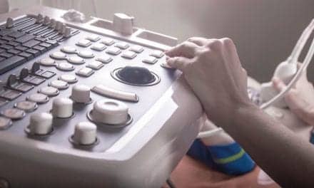
How It Works
The ABUS—a sonar device—works by sending sound waves into the breast. “They hit objects and send back echoes, which the computer converts to images,” Roux explained. The technology is a sound depiction of tissue’s characteristics, while mammograms are an x-ray depiction. “It looks at tissue from a different angle,” she said. “It is complementary to a mammogram, because with a mammogram cancer shows up white on a white background. In ultrasound, cancer shows up dark on a white background. With the contrast differences, characteristics of cancer can be picked up with sound.”
In addition, because it is a large format transducer, the ABUS can image a large span of the breast’s real estate on one picture. “You get a big field camera view versus a narrower focused view,” Roux said.
In addition to being a screening tool, Roux uses the ABUS for diagnosing patients with multiple cysts, multiple fibroadenomas, and other clinical complaints.
Reimbursement varies by state and insurer. Fortunately, “We haven’t had any trouble with patients being reimbursed for this type of study,” said Roux. “But the caveat is that you must tell patients that insurance will cover it, but they must meet their deductible first.”
From a usability perspective, “this machine is very technologist friendly and easy to use,” Roux said.
A Proven Commodity
Roux recalls asking patients if they wanted to volunteer for the new technology during the research period. Many were agreeable. After a few images, she recalls one patient who said she felt a little uncomfortable and wanted to discontinue.
“We were exceedingly lucky, because in those first two images cancer was discovered,” Roux said. “The patient was eternally grateful, as it may have saved her life.”
Roux says that most women find the ABUS technology to be comfortable. The ABUS has a patented Reverse Curve transducer that conforms to a woman’s anatomy, for better comfort and image performance. Furthermore, the system uses Compression Assist, a feature that applies light levels of compression automatically to the breast for increased ease and image reproducibility. Imaging takes about 15 minutes.
Roux will continue to value mammography in screening for cancer, because it is the only technology that can detect calcification—the earliest form of breast cancer. Mammography works well for women without dense breasts—that translates to about 60% of women.
But ultimately, Roux believes breast imagers should use a multimodality approach to screening including breast MRI for high-risk women. “Many different things work to help find cancer,” she said. “Know what to use when.”
References
- Boyd NF, Guo H, Martin LJ, et al. Mammographic density and the risk and detection of breast cancer. N Engl J Med. 2007;356:227-236.
- US Department of Health and Human Services. FDA. PMA P110006 summary of safety and effectiveness. September 2012.