Summary: Indiana University School of Medicine researchers used advanced PET imaging to identify a new genetic marker linked to Alzheimer’s disease progression, specifically targeting tau protein buildup, which could improve diagnostic tools and lead to new treatments.
Key Takeaways
- Researchers at Indiana University School of Medicine identified a new genetic marker (rs2113389) linked to tau protein buildup in Alzheimer’s disease using advanced PET imaging.
- The study is the largest to date, analyzing over 3,000 participants and showing this genetic variant explains more tau deposition than the APOE4 gene.
- The findings could improve early detection and lead to more targeted treatments, with plans for further validation in larger populations and lab models.
—————————————————————————————————————————————————————————
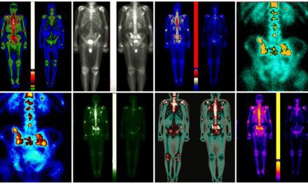
Indiana University School of Medicine researchers have made a significant discovery using advanced PET imaging to identify a new genetic marker involved in Alzheimer’s disease, which is marked by amyloid-beta plaques and tau tangles. Published in Nature Communications, the study’s findings could enhance diagnostic tools and lead to new treatment approaches.
The international team analyzed data from over 3,000 participants, making it the largest effort to date exploring the genetics behind PET-detected cortical tau. By using tau PET scans, they identified a specific genetic locus responsible for a significant portion of tau buildup in the brains of older adults at risk for Alzheimer’s.
Key Genetic Drivers of Alzheimer’s
“This novel genetic marker opens new research avenues by targeting the abnormal tau protein that forms tangles seen in Alzheimer’s,” says Andrew J. Saykin, PsyD, lead investigator and professor at IU School of Medicine.
This study differs from traditional genetic research on Alzheimer’s, which has primarily compared patients with amyloid plaques to healthy controls. By using tau PET scans as a continuous measurement, the team was able to detect genetic factors that drive tau accumulation. Saykin, who also directs the Indiana Alzheimer’s Disease Research Center, notes that while prior studies have identified genetic variants linked to amyloid buildup, finding the genetic causes behind tau deposition has been more challenging, mainly due to limited datasets with both PET and genetic information.
Their research fills that gap, demonstrating that a genetic variant called rs2113389, located between the genes CYP1B1 and RMDN2 on chromosome 2p22.2, is strongly associated with increased tau across multiple brain regions. This variant explains 4.3% of the variation in tau deposition, more than the well-known APOE4 gene, which accounts for 3.6%.
Targeted Alzheimer’s Treatments
“Further research is needed to understand what drives this genetic association,” Saykin says. The researchers plan to continue using tau PET imaging to explore this genetic locus and validate the findings in larger populations. Shannon L. Risacher, PhD, co-investigator, emphasizes the importance of expanding these studies globally, as most current data come from sites in the U.S., Canada, and Australia.
In addition to human studies, the team plans to use mouse models and lab-based systems to further validate these PET findings. “We want to replicate the effect in the lab to help advance drug development,” adds Kwangsik Nho, PhD, co-investigator.
Saykin highlights that combining advanced neuroimaging like PET with genetics is essential for understanding Alzheimer’s disease. “This approach could lead to earlier detection and more targeted treatments for those at risk,” he says.