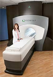
 While people born in the 1950s are beginning to close in on early retirement, nuclear medicine — born in the same era — is finding new ways to keep on working. Single photon emission computed tomography (SPECT) continues to evolve as a technology with expanding applications, and although considered mature, shows no signs of resting on its laurels or exiting the imaging scene any time soon.
Through three-dimensional tomographic images created by injecting radioisotopes that are distributed to specific organs within the body, physicians can analyze a study in multiple planes. Bone, kidney and lung scans remain major applications for SPECT imaging in providing significant information for finding bone tumors, determining kidney function and showing evidence of pulmonary emboli to the lungs. Count nuclear cardiology — with an estimated growth rate of 12 to 15 percent per year — and work involving brain imaging scans for studying seizure disorders and for the early diagnosis of neurodegenerative diseases (still a research tool) among SPECT applications garnering increasing attention.
SPECT’s growth in cardiology is moving the imaging method into the routine management and assessment of patients with coronary artery disease. And its ability to deliver functional information months before a problem can be detected in the brain with CT or MRI gives SPECT an imaging edge.
“The major strength of [SPECT] over the last eight years in terms of the research has been the prognostic and risk stratification application,” says James Udelson, M.D., associate chief of cardiology at New England Medical Center (Boston) and president of the American Society of Nuclear Cardiology (ASNC of Bethesda, Md.).
Please refer to the June 2001 issue for the complete story. For information on article reprints, contact Martin St. Denis


