MI readers pick the year’s most innovative products.

|
In considering the most innovative products of the past year, Medical Imaging looked to its readers. Based on their expressed interest and recommendations, the following are the products that were thought to be the most far-reaching, game-changing, or otherwise groundbreaking devices on the market.
Aquilion One: A Single Rotation
Diagnosing a stroke traditionally takes hours—sometimes even a full day—as patients are shuttled from test to test and then wait for their results. But the Aquilion One dynamic volume CT scanner from Toshiba, Tustin, Calif, allows clinicians to make a diagnosis within minutes.
At St Elizabeth Medical Center, Edgewood, Ky, patients who may have had a stroke are taken from the emergency department (ED) to the adjacent Aquilion One scanner, where clinicians perform a noncontrast head CT, start an IV, and do a brain-profusion study with the arteriogram. “We’re going from several hours to definitively diagnose a stroke to 10 minutes to diagnose a stroke,” says Jeff Dardinger, MD, director of imaging for the vascular institute. “And we’re getting much better pictures.”

|
| The Aquilion One from Toshiba images an entire organ within one gantry rotation. |
The Aquilion One, which was launched at RSNA 2007, is the culmination of 10 years of development and user feedback. The most exciting feature is the ability to image an entire organ, such as the heart or the brain, within one gantry rotation—an improvement over the company’s 256-row detector prototype.
“One of the major things we learned with the 256-row detector was it wasn’t wide enough to cover the entire brain and the entire heart, which is why we went to the 320-row detector that exists in the Aquilion One today,” said Doug Ryan, director of Toshiba’s CT Business Unit.
The 320-row detector covers 16 cm of anatomy, which reduces radiation and contrast dose as well as the exam time. “Now we can do the whole brain, and we can see the arteries really well, much better than we ever imagined,” Dardinger says. “We can actually do a CT venogram of the brain, which we were never able to do before.”
The technology also allows for better cardiac imaging. Although the hospital’s previous CT machine allowed for angiograms in 6 to 8 seconds, the heartbeat interfered with image quality. The Aquilion One has eliminated these misregistration problems. “We do the entire heart in a fifth of a second,” Dardinger said. “So, now there is no misregistration artifact at all. We’re seeing things much clearer and with much less distortion in literally less than a heartbeat.”
St Elizabeth acquired the scanner in August of this year, becoming the first community hospital in the United States to install the Aquilion One in an ED setting. Dardinger is excited about the unit’s contributions to better patient care. “With volume imaging, we’ll be able to do neuro imaging better, we’ll be able to do cardiac imaging better, and we’ll be able to do new techniques that we just couldn’t do with the 64-slice,” he said.
Artis Zeego: Precise Positioning
A little flexibility goes a long way toward improving imaging capabilities—especially in interventional radiology and cardiology. The Artis zeego from Siemens Medical Solutions, Malvern, Pa, features a robotic, multiaxis C-arm designed for virtually unrestricted positioning.
A physician can place the positioner at almost any point around the patient, allowing for complex movements such as tilted table scans in the peripherals. The isocenter position, which can be adjusted to the procedural needs or even the height of the physician, enables off-center rotational angiography for all areas of the body.
The adjustable isocenter also supports advanced 3D imaging techniques, including cross-sectional imaging through Siemens’ syngo DynaCT, which allows the physician to see the whole abdomen or the entire liver for chemoembolization and biopsies. The Artis zeego also offers views of the skull and the neck, and expanded views of the spine.
The robotic C-arm, which can move from one position to another within a fraction of a second, allows physicians to reproduce exact angulations without reinjecting contrast. The Artis zeego can be used for preoperative and postoperative imaging within the operating room.

|
| Philips’ Brilliance iCT provides improved image quality at a lower dose. |
Brilliance iCT: Improved Workflow
The CT scanner has become much more of a workhorse for hospitals in recent years. Once seen as a luxury item, this machine is now essential to diagnosis, treatment planning, and other key medical decisions in most facilities. So, it is no wonder that reliability and efficiency are of great importance to today’s medical imaging professionals.
“Dependability of that CT scanner is paramount,” says John Steidley, vice president of global CT marketing for Philips Healthcare, Andover, Mass. “It’s no longer a nice-to-have feature—it’s a necessary test, and it needs to be very dependable within the hospital.”
To meet these needs, Philips designed the 256-slice Brilliance iCT with a gantry that rotates four times per second. This means the unit can scan the heart within three heartbeats (5 to 8 seconds). The scanner is also designed to reduce radiation dosage and patient exposure.
The Brilliance iCT unit features an antiscatter grid called the nano-panel 2D. “It turns out 95% of the x-rays in a CT scan have scatter in the body, and rejecting that scatter information leads to better images,” Steidley says.
Of course, more slices mean more images, which can impact workflow. To help clinicians manage the images they receive, Philips has developed the Brilliance Everywhere thin client. The application allows for interdepartmental collaboration and can be accessed from any workstation or home office computer.
Steidley adds that the Brilliance iCT also allows easier and faster imaging for complicated cases, such as bariatric, pediatric, or cardiac patients. “We’ve really been able to deliver a CT that’s simple to use and provides improved image quality at a lower dose for a broad spectrum of cases,” he says.

|
| Carestream’s DRX-1 wireless DR detector is a portable and affordable solution. |
DRX-1: Portable Detector
Some clinicians long for digital imaging solutions, but budget constraints stand in their way. Others just don’t have the stomach for replacing their tried-and-true equipment with all-new machines. To address both of these concerns, Carestream Health, Rochester, NY, developed the DRX-1 wireless DR detector, which is the size of a cassette and can be used with existing wall-stand or table-based Buckys.
Without requiring any modifications to existing equipment, the 14- x 17-inch detector provides high-quality preview images within 5 seconds. Many users will already be familiar with the software interface. “When the image comes up on that console, it comes up in our direct-view software,” says Todd Minnigh, worldwide director of marketing, digital radiography, for Carestream Health. “If you already know how to use our CR or our DR, you already know how to use the DRX-1.”
The DRX-1 is set to launch in spring 2009, and Minnigh expects the wireless detector will greatly improve efficiency for facilities. “I think what you’ll see is places that implement this technology will have shorter wait times in the waiting room, and they’ll have capacity where they can do additional patients where they might not have been willing to schedule those patients so closely before,” he says.
He also believes it will improve patient care, as the technician will be able to perform the tests more quickly and without leaving the patient’s side. “Nobody enjoys getting x-rays,” Minnigh says. “They don’t want it to last 20 minutes; they’d like it to be done in 5 minutes. If you can do it quickly like that and stay with them the whole time, I think the patient experience will be much better.”

|
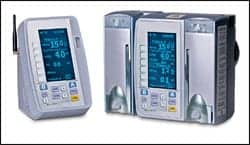
| MedRad’s Intego PET infusion system eliminates manual patient dose preparation, adjustment, and injection. |
Intego: Dose on Demand
The preparation and dispensing of radiopharmaceuticals for nuclear medicine has traditionally been a manual process.
“There’s a lot of handling, there’s a lot of inaccuracy, and whenever there’s a schedule problem—if the patient comes late or if the patient’s early—it’s much more difficult to respond to that because the radioactive tracer is always decaying,” says Alan Connor, marketing manager for MedRad, Warrendale, Pa.
On top of this, the short half-life of radiopharmaceuticals, such as FDG, often requires multiple deliveries per day. To improve the efficiency and safety of working with radiopharmaceuticals such as FDG, MedRad has created the Intego PET infusion system, which eliminates manual patient dose preparation, adjustment, and injection.
The Intego PET infusion system features automatic extraction of patient dose from a multidose vial, a saline test-inject feature, an automatic saline flush, and an air-detection system that disarms the system if air is present in the source administration set. An integrated ionization chamber measures each dose prior to injection, delivering it within 2% of the measured dose.
“Instead of having eight syringes for eight patients over a morning schedule, you’ll have one vial of FDG,” Connor says. “It will be contained inside the shielding of our Intego system, and then at any time during that morning, the technologist tells the system what dose to prepare. The system will prepare that dose and then deliver it to the patient.”
By eliminating the manual steps involved in preparing and injecting radiopharmaceuticals, the Intego system reduces radiation exposure to technicians by at least 20%. “We are basically bringing to the market a new way of handling these radioactive substances that is more precise, more flexible, and safer for the technologists who have to deliver that agent,” Connor says.
Mobile MIM: Images on the Go
Physicians may soon have images literally at their fingertips. Designed for Apple’s popular iPhone and iPod Touch, the Mobile MIM application from MIMVista Corp, Cleveland, will serve as a remote image viewer, allowing physicians and clinicians to access images when they are away from their workstations. The application is currently pending clearance from the FDA.
The alpha version, released earlier this year, provided multiplanar reconstruction of data sets from modalities including CT, PET, MRI, and SPECT, as well as multimodality image fusion. The multitouch interface of the iPhone allowed users to change image sets and planes as well as adjust zoom, fusion blending, and window/level.?
OsiriX: Open-Source Option
Generic software applications are slowly becoming a thing of the past as more specialized fields look to organize and access data in a specific way. This is certainly true of medical imaging, where precise image manipulation and integration tools are essential to both workflow and quality control.
Jumping on this trend is OsiriX imaging software, an open-source application developed by Osman Ratib, MD, PhD, FAHA, professor and chair of radiology of the Department of Medical Imaging and Information Sciences at the University Hospital of Geneva, along with Antoine Rosset, MD, and Joris Heiberger (see “Software Offers PET/CT/MR Image Fusion” from our July issue). The free application, which is designed for navigation and visualization of multimodality and multidimensional images, functions both as a DICOM PACS and as image-processing software.
OsiriX includes 2D Viewer, 3D Viewer, 4D Viewer, and 5D Viewer. The 3D Viewer offers all modern rendering modes, including multiplanar reconstruction, surface rendering, volume rendering, and maximum intensity projections. These modes support 4D data and are able to produce image fusion between two different series, such as PET/CT.
The software features ultrafast performance, an intuitive interactive user interface, and an open platform for the development of processing tools. OsiriX 3.3 was released in October and is compatible with Mac OS 10.5. An iPhone version of the software was announced at RSNA 2008.
ProSound 3500SX: New Features
The redesigned ProSound 3500SX 4D diagnostic ultrasound system from Aloka America Ltd, Wallingford, Conn, offers key features for the clinical applications of obstetrics and gynecology, breast, and urology. The system offers high-quality images and a customizable reporting package.
The redesign includes a flat-panel display and improved ergonomics. Enhanced 3D/4D imaging allows clinicians to save entire data sets to review later for faster exams. The system, which includes a USB port for media storage, features free 3D/4D viewing software for postprocessing on independent workstations. In addition, the ProSound 3500SX includes extended field of view to view the entire uterus for multiple gestations and placenta position.
Ann H. Carlson is a contributing writer for Medical Imaging. For more information, contact .


