
For pregnant women, ultrasound is a vital modality. But when it comes to a select population—obese pregnant women—ultrasound has limitations. Researchers, clinicians, and manufacturers take pause as they explore advancements and features for the future.
 |
Obesity in pregnant women presents a number of technical challenges for ultrasound clinicians, and with nearly one million overweight women in the United States delivering each year, the problems will not go away soon. Indeed, obesity has reached epidemic proportions with 66% of Americans overweight—including almost 5% considered morbidly obese—according to a Journal of the American Medical Association (JAMA) study published in 2006.1
In 1962, 13% of the American population was classified as obese. By 1994, this number had increased to 23%. Just 6 years later, this number had exceeded 30%. Now, confirmed by the JAMA study, being obese has officially become a marker for classifying pregnancy as high risk.
According to the New England Journal of Medicine, pregnant women who are overweight or obese have longer, more costly hospital stays and use more medical services than normal-weight women. Also, overweight women tend to visit the prenatal clinic fewer times than normal-weight women. Finally, higher rates of cesarean deliveries associated with obesity are increasingly responsible for longer hospital stays.
Information obtained by ultrasonography in obese pregnant women is often unsatisfactory because adipose tissue is a poor conductor of ultrasonic waves. Women who are obese during pregnancy run the risk of preeclampsia, a condition that causes high blood pressure, fluid retention, and swelling; gestational diabetes, cesarean section, and postpartum infection. Complications for the baby include macrosoma, which can impede labor and delivery; neural tube defects; and childhood obesity.
“Obesity is considered a clinical risk factor in obstetric practice that poses a particular challenge for the completion of the second-trimester fetal ultrasound survey,” said Phyllis Glanc, an assistant professor of medical imaging at the University of Toronto, in a report from the American Roentgen Ray Society (ARRS). Her investigators found that most pregnant women with a high body mass index had incomplete or unsatisfactory exams, and that more than one-quarter of them required additional ultrasound scans to complete their surveys. “Twenty-six percent of screenings in the study group were considered incomplete and required another scan, as compared with only 2.5% in the control group. This is a hot-button topic,” she said, “but we don’t believe it has been well documented.” Glanc presented her group’s results at the 2008 ARRS meeting in Washington, DC.
According to the study, the most common indications for recall were inability to visualize the fetus’ heart (16%), facial profile (10%), and spine (6%) compared with heart (0.5%) and spine (1%) in controls. Almost two-thirds of recalled exams remained categorized as having suboptimal visualization.
“One of the many challenges facing clinicians is the adaptive equipment necessary, including special beds and physical challenges for those caring for the patient,” said Loralei Thornburg, MD, Maternal Fetal Medicine, University of Rochester, Strong Memorial Hospital, Rochester, NY. “Also, because often these women are at increased risk for pregnancy complications, there is a need for additional monitoring and assessment of the pregnancy, which is more difficult to accomplish than in the nonobese patient.
“3D/4D ultrasound is starting to play an increasing role in evaluation of fetal anomalies and can help improve our assessment of fetal defects in utero,” she added. “However, these technologies are profoundly limited in obese women. For obese women, some experts are starting to advocate transvaginal ultrasound for fetal anatomy assessment, especially cardiac anatomy, earlier in pregnancy before transabdominal ultrasound is possible on morbidly obese patients, but this is still investigational. Some ultrasound manufacturers have also developed special transabdominal probes to help overcome the decreased penetration and resolution of traditional ultrasound probes in obese women. However, it is still not clear if any of these advances will be able to improve ultrasound resolution to equal that of the nonobese patient.”
“Ultrasound technology is improving, and this has improved visualization in obese pregnant women,” Thornburg continued. “However, one of the best and easiest tools available to clinicians to aid in ultrasound visualization in obese women is referral for ultrasound at the appropriate time in pregnancy. In general, ultrasound to evaluate fetal anatomy is more effective a few weeks later in obese patients. Additionally, using methods to project birth weight from an earlier gestational age before the fetus descends into the pelvis and the fluid volume decreases can make estimating fetal weight easier to obese pregnant women.”
A recent study published in Ultrasound in Obstetrics and Gynecology authored by Thornburg et al explores sonographic birth-weight prediction in obese patients using the gestation-adjusted prediction method (GAP).2 Thornburg demonstrated that the mean birth-weight error varied significantly between obese patients and normal-weight controls with birth weight underestimated in those patients with Class III obesity as compared to controls. The method predicts actual birth weight within 20% for all groups in more than 90% of cases. For all groups, the GAP method correctly excluded the presence of macrosomia with ≥90% specificity. Negative likelihood ratios for the prediction of macrosomia were between 0.4 and 0.6 for all groups, regardless of obesity. Conclusions: The GAP method of birth-weight prediction has been validated in women with gestational and pregestational diabetes as an accurate prediction method for birth-weight estimation. What is not known is whether the accuracy of this birth-weight prediction method extends to obese patients, especially those with morbid obesity in whom visualization may be severely limited.
Thornburg points out that, “One of the daily challenges that face clinicians and sonographers when working with obese pregnant patients is the additional time required and difficulty in fully assessing the fetus. Just because a patient is obese does not mean that her imaging will be worse than a nonobese patient because so much depends on how she carries the extra weight. A sonographer may have to be very creative in how they image the obese patient—using the umbilicus, placing the woman on her side, or raising the panus to view the lower uterus are just of the few of the ways that they can improve imaging—but there is significantly more work for the clinicians involved.”
Those patients who have both diabetes and obesity are probably the biggest challenge, since both not only increase the risk of pregnancy complications, but also adversely affect the ability to assess the fetus by ultrasound. Since these diseases often exist in tandem, these patients remain a key area for new improvements in ultrasound technology.
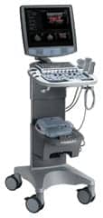 |
| Experts say sonographers may need to be creative when imaging an obese pregnant patient with ultrasound equipment. |
What’s Next?
“Unfortunately, there are limits to ultrasound technology and the penetration available,” Thornburg said. “I expect that we will see more use of early transvaginal ultrasound for fetal anatomy assessment. It is also likely that there will continue to be improvements in ultrasound probes and refining of ultrasound technology to optimize penetration and improve clarity of ultrasound in obese patients. However, it is still not clear if any of these changes will be able to improve outcomes for obese women in pregnancy, given the increased risk of pregnancy in obese women and the steadily increasing numbers of obese and morbidly obese women becoming pregnant.”
Lauren Dungy-Poythress, MD, medical director of Maternal Fetal Medicine, Community Health Network, Indianapolis, who is an ultrasound specialist, is an enthusiastic user of the Philips C5-1 PureWave transducer. She confirms the technical problems of ultrasound with obese pregnant women. “The bigger the woman is, the harder it is to get an accurate evaluation of the fetus,” she said. “But the new (C5-1) probe allows us to visualize the fetus better than ever. I’m excited to experience this level of detail and am very pleased with the quality of the image. It’s a godsend. It leads to a better prognosis and better care for the pregnant patient. It’s absolutely a great innovation. In the future I look for greater resolution, and this will allow better treatment.”
The use and development of the Philips C5-1 unit in difficult ultrasound cases is promising. In testing, the C5-1 unit demonstrated, among other advantages, reduced exam times anywhere from 2% to 38% in technically complicated cases; a reduction in pain and fatigue from scanning in 29% to 85% of the cases; a less need to push in order to achieve penetration of an organ or structure in 48% to 93% of the cases; and marked improvement in color sensitivity.
According to Philips, a main cause of ultrasound image degradation arises from the improper assumption of speed-of-sound characteristics when scanning patients with significant adipose layers. The resulting beam aberrations account for the loss of detail resolution seen in obese patients. For the first time, the C5-1 transducer works in conjunction with the iU22 to accommodate for the altered speed of high-frequency sound waves through adipose layers versus other tissue. In this unique mode, the ultrasound system becomes “aware” of increased adipose content and applies aberration-correction algorithms. The result is increased sharpness and clarity in image quality throughout the entire beam length.
Cathy Fix, RDMS, ultrasound supervisor at St Paul’s Hospital, Vancouver, British Columbia, Canada, in reference to ultrasound examination of obese patients using C5-1, said, “We increased diagnostic confidence for all our obstetrical exams ? especially with fetal heads and hearts ? lack of artifacts helps to increase our diagnostic confidence.”
The prevalence of obesity is a global problem that is rising at alarming rates, and it is a uniquely dangerous problem among pregnant women. Technology is trying to keep up with the health care hurdles this phenomenon engenders. Progress is being made, but researchers, clinicians, and manufacturers continue to tackle the challenges.
James Markland is a contributing writer for Medical Imaging. For more information, contact .
References
- Ogden CL, Carroll MD, Curtin LR, McDowell MA, Tabak CJ, Flegal KM. Prevalance of overweight and obesity in the United States, 1999-2004. JAMA. 2006;295:1549-1555.
- Thornburg LL, Barnes C, Glantz JC, Pressman EK. Sonographic birth-weight prediction in obese patients using the gestation-adjusted prediction method. Ultrasound Obstet Gynecol. 2008;32(1):66-70.

