A new study published in the European Heart Journal shows that CT can be a useful tool for making decisions regarding complex coronary disease treatment. Below, Scott Schubert, General Manager, Global Premium CT, GE Healthcare, sits down with AXIS Imaging News to discuss the study—the SYNTAX III Revolution trial—and reveals how GE Healthcare played an integral role in it.
AXIS Imaging News: Please explain a little bit about the trial and its findings.

Scott Schubert
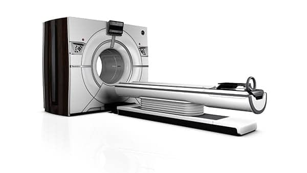
Scott Schubert: The SYNTAX III Revolution trial randomized two heart teams—each made up of an interventional cardiologist, cardiac surgeon, and radiologist—to make treatment recommendations for 223 patients with diagnosed left main or three vessel disease. One team reviewed data from GE Healthcare’s high-definition Revolution CT scanner with a fractional flow reserve (FFR) CT assessment to make their treatment recommendations. The second team used information from the conventional coronary cine-angiogram to make a treatment recommendation.
The study showed “almost perfect agreement” (Cohen’s kappa of 0.82) in the team’s treatment recommendations, suggesting that the less-invasive CT exam may be just as informative as the invasive cine-angiography.
AXIS: Why are these findings so noteworthy?
Schubert: In the United States, CAD is the leading cause of death for both men and women. It’s estimated that half of healthy 40-year-old males and one in three healthy 40-year-old women will eventually develop the disease.
With these rising numbers, effectively diagnosing and treating CAD is a major focus in cardiovascular research, and one in which CT has played a larger and larger role. In patients with complex, severe CAD, invasive cine-angiography is used to visualize the coronary anatomy and guide treatment decisions. But this study suggests that a simple CT scan may be just as effective, and far less invasive. This is significant, since it could offer patients more comfortable experiences while still providing heart teams with detailed anatomical information.
In a survey of the surgeons involved in the trial, 84% said they could even do surgery based on the CT scan alone. These results speak to the level of image detail and amount of information provided by the CT scan and highlight the potential of this technology to not only diagnose complex coronary disease, but also assist in subsequent heart team decision-making.
AXIS: What could this mean for cardiac teams (and patients) moving forward?
Schubert: These findings suggest an opportunity to streamline the care pathway. The multi-slice CT scan offers an easier, less-invasive patient experience; takes less time; and a provides a clear, comprehensive anatomical picture to assist clinicians with diagnoses and treatment recommendations.
Professor Patrick W. Serruys, principal investigator and study chairman at Cardialysis, even suggests the CT’s potential as a diagnostic tool for complex coronary disease could enable so-called ‘cath labs’ to transition from diagnostic to interventional suites that concentrate on the significant load of coronary and structural heart disease patients.
AXIS: The trial identifies CT as a non-invasive diagnostic alternative to coronary angiography for complex coronary disease. What are the differences between these two procedures?
Schubert: Coronary angiograms are the current industry standard to help clinicians diagnose heart disease and identify the best course of treatment. During these procedures, a small incision is made to insert a catheter, which is used to inject dye into the blood vessels of patients’ hearts. A special x-ray machine is then used to image the dye’s movement through patients’ blood vessels. Should a blockage be found, clinicians may open patients’ clogged heart arteries (angioplasty) during the exam. The procedure usually takes about an hour.
CT scans also use advanced x-ray technology to image the body from different angles to produce tomographic, highly detailed images of internal organs, bones, soft tissue and blood vessels. The tomographic images gathered during the scan can then be reformatted in multiple planes for clinicians to observe anatomic abnormalities—or in this case, artery blockages.
Both procedures emit radiation, but according to the Syntax III trial results, the radiation dose (effective dose) in the CT in this study was about 50% lower than the dose of the conventional angiography.
AXIS: What else do you want AXIS Imaging News readers to know about this study and its findings?
Schubert: Cardiovascular disease affects millions of patients globally, and our goal is to improve access to high-quality imaging solutions by providing cost-effective tools that help physicians confidently and efficiently diagnose and treat these patients. This study highlights the potential of this technology to greatly improve the care pathway as well as patient care using noninvasive techniques.
The next step will be an outcomes trial of 100 patients to see what happens when the heart teams follow through with their treatment decisions based on either the CT scan with FFR or the coronary cine-angiography. This trial will again be conducted by Cardialysis on behalf of the European Cardiovascular Research Institute.
