Manufacturers are stepping up safety efforts with better dose control, improved delivery systems, and stronger educational efforts about contrast protocols.
 |
Contrast agents have long been essential diagnostic tools for radiologists, as they help provide the crystal-clear images required to make accurate diagnoses. But the use of these agents is not without risk.
The main concern is patient safety. Administering the wrong contrast agent or delivering the wrong dose can cause serious side effects. For example, a rare but severe condition called nephrogenic systemic fibrosis (NSF) has been linked to the use of gadolinium-based contrast agents.
According to a recent study published in the American Journal of Roentgenology, adverse reactions to contrast agents are rare (See our recent news story). But addressing safety concerns continues to be a high priority for the imaging industry, which is pushing for better dose control, improved delivery systems, and stronger education efforts about contrast safety protocols.
Ultimately, it is up to physicians and radiology technicians to ensure that patients receive the right dose of the contrast agents they need. But manufacturers of contrast agents and delivery systems are also taking steps to reduce the potential for error when health care professionals use their products.
“Delivering the right contrast agent to the right patient in the right concentration is very important in the radiology environment,” said Rhonda Soest, RN, marketing director for the pharmaceuticals division of Covidien, Hazelwood, Mo. “We’re always working toward solutions that help reduce risks associated with the delivery of contrast.”
The RFID Advantage
To promote patient safety as well as streamline workflow for radiology departments, Covidien launched a delivery system in July 2008 that incorporates radio-frequency identification (RFID).
“It’s really the next-generation delivery solution,” Soest said.
The Optivantage dual-head CT injector with RFID interfaces with the company’s Ultraject prefilled syringes, which feature embedded RFID tags. When the syringe is placed in the injector, the syringe’s RFID tag transmits to the injector, which records the volume, the lot number, and the concentration of the contrast agent. The system also generates a printed label that clinicians can add to the patient chart without the need for manual notation.
“It helps ensure that the correct information is transcribed to the patient chart,” Soest said. “It really helps improve efficiency and accuracy.”
The combined use of the RFID injector and the RFID-enabled syringe was designed to help eliminate?many?of the risks associated with unlabeled syringes, prevent cross-contamination, and avoid accidental emboli.
“It will alert you if the syringe has been used or is empty so that you can prevent?air?emboli,” Soest said. “So, the label really creates an intelligent interface between the Ultraject?prefilled syringe and the Optivantage dual-head injector.”
Soest notes that Covidien continually assesses how the company’s delivery systems match up to patient safety guidelines and recommendations from organizations such as The Joint Commission. For example, the Ultraject prefilled syringes are designed to comply with The Joint Commission’s recommendation that physicians use the most ready-to-use form of the?drug?available.
“When you combine that with RFID, not only does that meet The Joint Commission recommendations and standards, but it also reduces the need for hand labeling,” Soest said. “It decreases the number of steps necessary for radiology departments to be compliant with these national patient safety guidelines.”
Patency Check
 |
| Mark Hibbard, MD, PhD |
In July 2008, Covidien also introduced the Optistar Elite injector for MR imaging. The most significant feature of this injector is the Patency Check function, which confirms that the patient’s vein is open before the contrast agent is delivered.
Ruling out any problems with the vein is imperative to both maintaining the patient’s safety and ensuring a successful diagnostic exam. The preprogrammed?Patency Check feature slowly delivers 10 mL of saline into the vein to aid clinicians in the prevention of extravasation, so that the saline does not leak into the surrounding tissue.
“The Optistar Elite also has a visual alert on the injector powerhead and on the control console that will warn you if the programmed pressure limit is too high,” Soest said. “And if there is a blockage, the alarm will engage to let you know that the injector is detecting it—just in case you didn’t?visually note?that blockage or extravasation.”
To keep physicians abreast of the appropriate ways to use Covidien’s contrast agents and delivery systems, the company educates customers both through its sales organization and at the presentations it hosts during the RSNA annual meeting. At RSNA 2009, for example, Covidien’s chief medical officer and vice president of medical affairs presented on the latest information about NSF and contrast use.
“On both the CT and the MR side of the house, the risks associated with contrast and contrast administration are always a concern,” Soest said. “Our job is to make sure that we educate physicians and continuously work toward solutions that are designed to help reduce those risks.”
Contrast Development
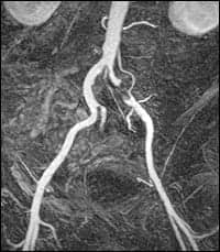 |
| ABLAVARTM enhanced angiography, first-pass image. |
One of the keys to using contrast agents safely is making sure to administer the appropriate agent for the exam being performed. On the manufacturing side, this can mean developing new contrast agents for different medical applications.
For example, Lantheus Medical Imaging Inc, North Billerica, Mass, recently launched ABLAVAR (gadofosveset trisodium), the first FDA-approved injectable contrast imaging agent for MRA. ABLAVAR is indicated for evaluating aortoiliac occlusive disease (AIOD) in adults with known or suspected peripheral vascular disease (PVD).
“We knew it was a serious medical condition that needed to be properly diagnosed so radiologists and surgeons could manage their patients effectively and relieve them of the severe pain and suffering associated with PVD,” said Mark Hibberd, MD, PhD, senior medical director of global medical affairs for Lantheus Medical Imaging.
When developing a new contrast agent, determining the right dose is crucial to patient safety. The key is finding the minimum effective dose so that the image quality is still high, but patient exposure is as low as possible. To this end, ABLAVAR is administered in a single injection at the lowest dose of any existing agent, notes Hibberd.
“With all drugs, the higher the dose, the more likely it is to cause complications,” he said. “In some situations, you would end up giving perhaps one tenth of the dose of gadolinium with ABLAVAR as you would with some other agents. That is a significant safety advantage.”
ABLAVAR is also a blood-pool contrast imaging agent, which means it binds to the serum albumin in the blood. This is an advantage over extracellular contrast agents that tend to leak out of the bloodstream more quickly—meaning that if the images aren’t successfully captured the first time, patients have to receive a second dose to repeat the test.
Instead, ABLAVAR circulates with the blood, which buys clinicians more time to perform the exam. “You actually have up to an hour to do your imaging,” said Hibberd, adding that ABLAVAR is currently the only gadolinium-based blood-pool contrast agent for MRI available. “Since the imaging usually takes only 30 seconds to a few minutes, if anything goes wrong, you can do it over pretty easily.”
Hibberd credits this as well as ABLAVAR’s low dose with the fact that although 90,000 doses of the gadolinium-based contrast agent have been made available, no incidences of NSF have occurred.
“We haven’t seen any sign of that particularly unusual side effect,” he said.
Contrast Versus Noncontrast
When weighing the pros and cons of using a contrast agent for a diagnostic exam, it is also important to consider the safety concerns of noncontrast alternatives.
For example, the ABLAVAR contrast agent was developed to create a safer option to detect AIOD than x-ray angiography, a relatively long and invasive procedure that can be uncomfortable for patients. X-ray angiography involves a significant dose of ionizing radiation and requires inserting catheters into the patient’s arteries, which creates a risk for tearing the arteries or even infection. Using a contrast agent with MRA, on the other hand, is a much quicker and minimally invasive procedure.
“We really liked the idea of developing something that could replace x-ray angiography for diagnostic purposes, was minimally invasive, and required only a short intravenous injection,” Hibberd said. “Also, MR imaging doesn’t contain ionizing radiation and doesn’t add any burden to the radiation doses that patients achieve over a lifetime.”
In clinical trials, the accuracy of the exam results with ABLAVAR proved comparable to those with x-ray angiography, which is the gold standard for detecting AIOD. In addition, MRA exams using the ABLAVAR contrast agent were significantly superior for AIOD detection than noncontrast MRA.
When considering all of these factors, Hibberd notes that using ABLAVAR in conjunction with MRA has several advantages for patients over x-ray angiography.
“When it comes down to patient care, it’s always appropriate to balance the risks with the benefits associated with using drugs and also contrast agents,” Hibberd said. “So, if you have the right test for the right diagnosis that you want to make or exclude, then that’s a good place to start. And if you’re sure that you’re using the correct and lowest possible dose, then I think you’re in a good place from a practice point of view.”
Ann H. Carlson is a contributing writer for Axis Imaging News.
INFORMATICS TOOL REDUCES ERRORS
MEDRAD’s Certegra Informatics Platform helps automate, manage, and record contrast data for imaging facilities
 |
| Connect.PACS immediately transfers a patient?s contrast-injection record to PACS and eliminates the need to manually enter injection information. |
When it comes to working with contrast agents, details matter. Patient safety depends on precision to ensure that the correct dosage is administered at the optimal time to the right patient.
But due to the sheer volume of imaging exams today, details can get lost in the shuffle. Unintentional errors, such as accidentally copying the wrong dosage amount into a patient’s chart, can have severe consequences for patient safety.
Managing and standardizing the data collection for these procedures can go a long way toward reducing the potential for error—with the added bonus of streamlining workflow for imaging facilities. Enter the Certegra Informatics Platform from MEDRAD Inc, Warrendale, Pa, which includes features to help automate, manage, and store contrast data.
“When you’re writing down numbers, leaving Post-its here and there, and you have to remember to go back and write something down, the opportunity for error exists,” said Anthony Cinalli, executive director of MEDRAD Radiology. “So, by having all of that information automatically stored and available, you’re removing variables associated with error potential.”
In 2008, MEDRAD introduced Certegra’s P3T (Personalized Patient Protocol Technology) software, which incorporates an FDA-cleared algorithm to automatically tailor the contrast dose to each patient. The algorithm takes into account factors such as the patient’s weight and medical history as well as the procedure itself to recommend the contrast protocol for that patient.
“You’re really defining the amount of contrast and the flow rate that you’re going to need to perform that injection,” Cinalli said. “By recommending the optimal dose to give to the patient, you avoid excessive contrast being delivered.”
P3T also recommends the optimal time to take the scan so that imaging professionals can be sure that the contrast agent is in the region of interest during the actual imaging.
P3T Cardiac is available for cardiac CT, CT angiography, and pulmonary embolism applications, while P3T Abdomen is used for dynamic liver imaging and imaging of the pancreas and kidneys. The idea is to take the guesswork out of contrast dosage for imaging professionals.
“Our goal is to take that variable out and say it’s not a matter of what works best most of the time; we’ll create the optimal protocol all of the time,” Cinalli said.
The specifics of the exam, such as the flow rates and dosage used, are automatically recorded and made available to radiologists at their PACS viewing stations through Certegra’s Connect.PACS application. This allows radiologists to view this data on the screen at the same time that they are reading the study, rather than having to separately track down important information about adverse reactions or pressure limits associated with the procedure.
“All of the information is available at their fingertips with Connect.PACS in their PACS viewing station,” said Cinalli.
The Certegra platform also includes the Manage.Report software application, which gives administrators the opportunity to analyze contrast data associated with multiple patients. This can be used to answer a variety of questions, such as how many patients came in for a particular study that week, how much contrast was used overall, and what types of complications arose.
Organizing and centralizing these types of details can help imaging centers optimize processes such as inventory reordering. By determining how much contrast is used during a given time period, for example, imaging centers can better predict how much they will use in a year.
“By having all of this data in one central location, you actually open up analysis to other areas and departments within the hospital environment,” Cinalli said. “You can really start to optimize your workflow from an efficiency standpoint.”
—Ann H. Carlson