Cardiac CTA is fast approaching angiography as a diagnostic tool. But in setting up advanced cardiac imaging services, how do you avoid battles between cardiologists and radiologists?
 |
| Many professionals agree that collaboration between radiologists and cardiologists is the key to successful cardiac CTA. |
With more than 79 million Americans suffering from cardiovascular disease, cardiac imaging is one of the fastest-growing areas in medical imaging. “The cardiovascular system is the most commonly imaged organ system,” said Alan D. Kaye, MD, FACR, with Advanced Radiology Consultants in Trumbull, Conn. The advent of coronary CTA [CT angiography] and other cross-sectional cardiac imaging techniques is expected to have a huge effect on the workup of patients with cardiovascular disease, a process that is currently evolving.
“Cardiovascular disease is on the rise, and it is going to be critical to have a convenient, cost-effective, and accurate means of testing. CTA offers that,” said Todd Kranpitz, CRA, MS, director of imaging services at King’s Daughters Medical Center in Ashland, Ky.
Though the procedure’s acceptance grows steadily, there are some challenges to implementing advanced cardiac services, which can include procuring the resources and establishing processes. Some facilities struggle with the competition between radiology and cardiology specialties, although many agree the ideal methodology involves collaboration. “Radiologists are not willing to give up the ability to read the coronary CTAs, cardiologists want to be involved, and it is playing out differently in individual locations around the country,” said Kaye.
The result is often market dependent. “The Healthcare Advisory Board conducted a poll that found that the most common way to perform cardiac CTA was through collaboration with both radiologists and cardiologists,” said Kaye, noting that all three models (collaboration, radiologists reading alone, and cardiologists reading alone) were in use. “It’s market dependent, but most people agree that the best way to do it is to collaborate,” said Kaye.
Middle Ground
Collaboration is the method employed at King’s Daughters, but though the transition was smoother than in some instances, there were challenges. “Because we have a cooperative vascular service, there were many shared resources already, such as information service personnel. So on the technical side, it worked out extremely well,” said Kranpitz.
Kim Grooms, RN, nurse manager of Invasive Cardiac and Vascular Services at King’s Daughters, agrees with Kranpitz, adding, “We worked through it and have not heard any negatives from either group of doctors,” said Grooms.
Physicians from both groups perform the readings. The most common procedure is for the cardiologist to read the heart vessels and the radiologist to “over-read” the image, looking for other pathology. “Numerous studies have shown a significant percentage of incidental yet clinically significant findings on the exams that are anatomically separate from the CT arteriogram portion of the test,” said Kaye.
Kranpitz estimates that approximately 20% of all heart patients have pathology outside of the heart, meaning that if a cardiologist alone reads the exams, there could be as many as one in five patients misdiagnosed. Because radiologists are the only ones qualified to interpret the entire image, radiologist involvement is required to reduce liability as well as the risk of misdiagnosis.
A joint effort, however, faces more obstacles than getting the two groups to work together. Billing and reimbursement issues must be worked out. “It is not clear yet what the appropriate way is for two different physicians to be compensated for interpreting different aspects of the same examination,” said Kaye.
The “curve ball,” according to Kranpitz, is that whoever bills for the procedure is also liable. So cardiologists need the radiologists to read the complete exam to reduce their risk, but radiologists also need to review the cardiac vessels to protect themselves.
No one has yet developed an ideal solution. “At a national conference I recently attended, we had a roundtable discussion with eight people from eight different facilities, and we had eight different arrangements suggested,” said Kranpitz, adding that no one solution was agreed upon.
Staking Ground
Cardiologists claim, according to Kaye, that they are the only ones who know the heart and are therefore qualified to read the related images. But Kaye feels the heart is like any other organ. “A cardiologist is not required to interpret a coronary CTA any more than a neurosurgeon is needed to interpret a brain CTA, a urologist to interpret a kidney CT scan, or an OB/GYN to interpret a mammogram,” he said.
 |
| One expert estimates that 20% of all heart patients have pathology outside of the heart, making a CTA read by a radiologist vital. |
Some of the debate is related to economics. Kaye suggests that cardiology practices are cautious about ceding control of the imaging procedure to radiology because of the potential financial impact. “Imaging has become a mainstay of cardiology practice, and cardiologists self-refer a tremendous amount of imaging,” said Kaye.
Their control of the diagnostic workup, and thereby the demand for the examination, means they can confidently make the investment needed to add advanced cardiac imaging services. Kranpitz estimates that implementing these programs can cost $2 million, and that is conservative. But the return can take a while to realize. Kranpitz estimates that about five procedures must be completed per day to break even, and even then the payback would be 20 years out. “This is a sharp contrast to nuclear imaging, where the investment is lower and the reimbursement higher,” said Kranpitz.
The technology, however, can be used to perform CTAs on other body parts, such as the carotid and pulmonary regions. At King’s Daughters, the team performs approximately 3,500 to 4,000 CT procedures per month, 300 of which are CTAs. “CT angiographies of other parts of the body are every bit as critical as CTA of the heart for all of the same reasons,” said Kranpitz.
The procedure is noninvasive and requires a short patient recovery only if beta blockers, other medications, or contrasts have been used. The workstation permits 3D reconstruction (“an absolute dream for presurgical planning,” said Kranpitz), and PACS permits the images to be sent to physicians at remote locations.
“The literature has reached the tipping point where the coronary CTA clearly has some important uses, but many factors impact its adoption,” said Kaye, noting that the “turf war” and economics are some of the more important factors.
Giving Ground
Cardiologists, Kaye suggests, are more likely to support the technology when they play a role, and their caution may be well founded. Coronary CTA is poised to replace diagnostic catheterization. “Coronary catheterization will eventually be reserved for patients with positive coronary CTA or who have a very high likelihood of disease, based on clinical parameters, that will require intervention,” said Kaye.
Cardiac CTA may also replace nuclear cardiac exams. “Coronary CTA has the potential to replace nuclear SPECT because the latter test provides an indirect way of determining whether there is narrowing in the arteries. However, some argue that as a functional test, it is still a valuable exam. The algorithm hasn’t been worked out yet, but coronary CTA will undoubtedly replace at least some nuclear SPECT exams,” said Kaye.
“There is a comparison between performing nuclear cardiology exams and coronary CTA,” concurs Kranpitz, who notes the two are opposite in most requirements, such as space, operation, and maintenance. But despite the competition, cardiologists are interested in the modality.
King’s Daughters marketed the new service to its physicians and found excitement. Kranpitz recalls that two cardiologists chose to attend a weeklong training session so they could incorporate the service into their busy practices.
Imaging centers and radiologists may also choose to market the service directly to primary care physicians. Kaye notes some radiologists have developed very successful coronary CTA programs using this method. “Primary care physicians view this as an asset because they maintain control of the patient. Many feel that the high negative predictive value of the exam may obviate the need to refer to a cardiologist,” said Kaye.
 |
| King’s Daughters Medical Center in Ashland, Ky. |
Despite the value of coronary CTA, however, its use should be applied intelligently. The exam is not without hazards. Radiation exposure, particularly for young women, presents risks as does the use of contrast agents, notes Kranpitz. “Just because you are able to do a test does not mean it is the right test. We have to account for its effectiveness and ability to improve patient care,” said Kranpitz.
The American College of Radiology has issued a Practice Guideline on cardiac CT that specifically includes components relating to CTA, and the American College of Cardiology Foundation (ACCF of Washington, DC) released related guidelines last year. The Appropriateness Criteria for CCT and CMR were developed in collaboration with the American College of Radiology, Society of Cardiovascular Computed Tomography, Society for Cardiovascular Magnetic Resonance, American Society of Nuclear Cardiology, North American Society for Cardiac Imaging, Society for Cardiovascular Angiography and Interventions, and Society of Interventional Radiology. The document reviews the risks and benefits associated with various procedures and makes appropriate recommendations. The goal is to help patients bare their hearts, not break them.
Renee DiIulio is a contributing writer for Axis Imaging News. For more information, contact .
The Basics for a Cardiac CTA Program
A cardiac CTA cannot be performed without a physician to read the exam, but it also cannot be performed without the proper equipment, space, and personnel in place, all of which perform critical roles.
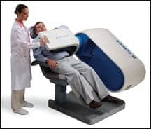
One of the first pieces to a cardiac CTA program is the equipment itself. Most programs, such as King’s Daughters Medical Center of Ashland, Ky, use a 64-slice CT machine. Slices, however, are not the best way to measure capability, said Alan D. Kaye, MD, FACR, with Advanced Radiology Consultants in Trumbull, Conn. “The number of slices is an inappropriate benchmark to use. It’s really the spatial and temporal resolution that needs to be sufficient,” said Kaye. The technology must permit images to show small details and freeze the motion of the heart. Kaye estimates that the machines with this technology cost approximately $1 million and more.
Todd Kranpitz, CRA, MS, director of imaging services at King’s Daughters, suggests the best way to determine the effectiveness of the technology is to do the homework. He suggests that site visits last longer than 30 minutes and involve those who will actually perform the procedure. “You want to sit down and review all of the issues that are important,” said Kranpitz.
Once the equipment is available, it needs a home, one with proper space and shielding. This room may include a power injector as well as a dedicated chiller; space will also be needed for patient recovery, whether in the unit or elsewhere.
To interpret the exams, the facility will need a workstation, where the images can be processed, and a display station on which physicians can review the exam. Adequate data lines are needed to transfer the data, and image archives are needed for storage. “The exams are very data intensive. It takes a tremendous computer to reconstruct the images, ” said Kranpitz, who adds that it also takes significant skill.
“To some degree, the procedure is operator-dependent. It takes a fair amount of skill to know the software, the anatomy, and how to process the images,” said Kranpitz. He hired an experienced professional to perform this work with positive results. “When we first started performing cardiac CTA, it took 50 minutes just to process the data, but we have now reduced that to 15 to 20 minutes,” said Kranpitz. The entire exam takes about 35 minutes; from the patient perspective, it takes an hour to an hour and a half, depending on the recovery time needed.
A nurse or other suitable professional who can monitor the patient during recovery as well as the exam itself (ie, physician or nurse practitioner) rounds out the care team. Proper education and training assure everyone knows their role. “Implementing a cardiac CTA program is a huge commitment,” said Kranpitz, citing education, staffing, and technology as important variables.
—R. DiIulio