 |
MRI |
New Applications: As Easy as CZT!
Promoting PET for Better Outcomes
New Applications: As Easy as CZT!
Spectrum Dynamics, based in Caesarea, Israel, with sales offices in Danville, Calif, reports that it now has installed 25 of its Cadmium-Zinc-Telluride (CZT)-based D-SPECT systems and that orders continue to grow as other imaging companies turn to this new type of gamma camera technology.
 |
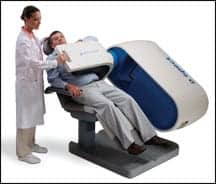
| The D-SPECT Cardiac Imaging System, powered by BroadView Technology, reduces the time of acquisition for a gated SPECT study to as little as 2 minutes. |
The D-SPECT? Cardiac Imaging System, powered by BroadView? Technology, significantly reduces the time of acquisition for a gated SPECT study to as little as 2 minutes, thereby improving throughput, as well as providing clinics a smaller device footprint.
Josh Gurewitz, vice president of sales and marketing at Spectrum Dynamics, says that the biggest advantage to this CZT-based camera is that its energy resolution is twice as good as the older sodium iodide technology.
He said, “The ability to discriminate different isotopes at the same time is much better with CZT. The problem in the past with CZT was the cost. It’s an expensive material and supply was limited in the past. But with companies like Spectrum Dynamics using it since 2006 and a major competitor announcing that their new product will be based on CZT, we’re seeing the supply improve substantially and the cost has come down.”
Since its introduction in 2006, the company has published research and trials benchmarking its D-SPECT system against traditional SPECT cardiology systems.
In May 2009, a performance evaluation was published in the journal, Physics in Medicine and Biology. The abstract concluded that “The D-SPECT has superior sensitivity to that of a conventional system with similar spatial resolution. It also has excellent energy resolution and count rate characteristics, which should prove useful in dynamic and dual radionuclide studies.”1
The results of another study published this year in the Journal of Nuclear Medicine showed that “the new camera had a count sensitivity that was 10 times higher than that of the conventional camera and a compensated spatial resolution that was moderately better.”2
The same abstract also indicated that a D-SPECT’s faster 2-minute studies compared well with 11-minute studies, showing “good-to-excellent image quality and improved myocardial edge definition for the new camera.”2
Gurewitz also added that CZT-based SPECT technology provides great promise for new techniques. He said, “There are new applications that are possible with these new technologies, such as being able to provide the cardiologist with flow data. That’s something that the old sodium iodide systems will just never be able to do. Also, the ability to look at multiple isotopes simultaneously because of the improved energy resolution of CZT is another future application.”
In addition to the faster throughput, the D-SPECT system features a unique seating design for improved patient comfort. An open chair allows patients to remain seated upright without the need to have an arm raised over the chest, as with more traditional SPECT systems.
—Tor Valenza
References
- Erlandsson K, Kacperski K, Van Gramberg D, Hutton BF. Performance evaluation of D-SPECT: a novel SPECT system for nuclear cardiology. Phys Med Biol. 54;9:2635-2649 (www.iop.org/EJ/abstract/0031-9155/54/9/003/. Accessed on July 28, 2009.
- Gambhir S, Berman D, Ziffer J, et al. A novel high-sensitivity rapid-acquisition single-photon cardiac imaging camera. Nucl Med. 2009;50:635-643 (jnm.snmjournals.org/cgi/content/abstract/50/4/635). Accessed on July 28, 2009.
Promoting PET for Better Outcomes
The Institute for Molecular Technologies (IMT), a council of the Academy of Molecular Imaging (AMI), has introduced a program to generate awareness and enhance the quality of positron emission tomography (PET) imaging. Called ACETM for Awareness, Communication, and Education, the program combines educational and clinical resources in easy-to-use kits designed to educate and improve the skills of interpreting physicians. There are separate resource kits for oncologic and cardiac PET applications.
PET is a powerful imaging modality that enables physicians to detect and stage many types of cancer quickly and accurately. If PET/CT is performed, a CT scan is obtained and the PET and CT images are co-registered. The results are 3D images of the body that can be used to localize abnormal processes with great precision. This combination of physiologic and anatomic information yields information that is unobtainable by other imaging modalities.
PET and PET/CT studies are convenient and easy for the patient. Scanning begins approximately 1 hour after the injection of the PET tracer (usually a fluorine-based glucose analog) to allow sufficient time for the tracer to accumulate in abnormal tissue. While lying down, the patient moves through the scanner to obtain images of the body. The imaging procedure usually takes 20 to 30 minutes, but may take longer if more of the body is imaged.
According to AMI, PET/CT is used to differentiate malignant from benign lesions, such as pulmonary nodules. However, it is more commonly used for initial staging of patients with known cancer, restaging after therapy, and detection of recurrence. The use of PET/CT in therapy monitoring is increasing, particularly in patients being treated for lymphoma.
“Imaging centers and physicians will find in ACE a valuable set of tools to educate referring physicians on the proper use and value of PET imaging,” said IMT chair Richard Frank, MD, PhD. “Even more importantly, ACE will ultimately make PET imaging more accessible to patients who need accurate information about their cancer or heart disease.”
Gail Rodriquez, AMI’s business development executive, said, “We are concerned about the appropriate use of PET technology, that is, when to use it and how to use it. We believe that PET is far too underutilized for cardiology and oncology applications. “
ACE provides turnkey assistance to PET imaging departments and physicians who wish to improve and promote their imaging services.
Users of the ACE Referral Education Kit will be able to raise awareness among their referring physicians about the clinical value of PET imaging utilizing easily customizable materials such as program announcement templates, imaging algorithms, sample press releases, case studies, clinical reprints, and patient education brochures. Components of the ACE Interpretation Resource Kit include protocols, case studies, clinical reprints, coding and reimbursement assistance, dictation pointers, and sample reports. All of these resources are intended to enhance imaging providers’ service quality and reports.
As a key example of the efficacy of PET scanning, Rodriquez pointed to a study recently published in the Journal of Clinical Oncology. “PET scan results led to a change in patients’ management in 36.5% of cases,” she said. “What we want to say is here’s how to use PET. We give them tools, clinical reprints, and the best ways to use PET effectively.”
The educational messages in ACE are based on the most up-to-date clinical data and practice guidelines and conform to all compliance and legal standards. Its modular framework is designed to accommodate new information and improvements as they become available, and offers a means to update the kits as other indications and modalities gain regulatory approval.
“The Academy of Molecular Imaging is pleased to offer this unique and valuable resource to the molecular imaging community,” said AMI president Tim McCarthy, PhD. “ACE demonstrates AMI’s commitment to be at the vanguard of molecular imaging and is an excellent example of how science and clinical practice can work together to ensure that patients receive the appropriate test at the appropriate time.”
—James Markland