A new ultrasound technology called Shear Wave Elastography promises to improve cost-effectiveness and enhance clinical workflow.
A new ultrasound technology called Shear Wave Elastography has been developed that provides not only exceptional gray scale or B-mode imaging, but quantitative real-time ultrasound elastography as well. Shear Wave Elastography adds important information by quantitatively measuring soft tissue stiffness in real time.1 The technique can capture ultrasound shear wave propagation in tissue, which was, until now, undetectable. The speed of the shear wave is directly related to the stiffness of the tissue. By generating, capturing, and quantifying shear wave propagation speed, this technology can produce an objective assessment of tissue stiffness or elasticity. Knowing tissue elasticity is an important step in the diagnostic process as tissue elasticity is related to pathology.
Shear Wave Elastography is regarded by many practitioners and sonographers as the next generation of ultrasound technology, which promises significant clinical and imaging management benefits. It has followed major advances, all of which have increased imaging specificity such as color Doppler, harmonics, and compound imaging, and is expected by many to be adopted in ultrasound systems as a key technology alongside these predecessors. Its clinical and economic value cannot be underestimated.
Using Shear Waves to Identify Tissue Stiffness
Shear waves are waves that move in a perpendicular manner to the direction of the tissue shear displacement. Shear waves travel faster in harder tissues. Shear Wave Elastography generates low-frequency shear waves in the body by employing focalized beams of acoustic energy using conventional transducers. As shear waves travel through the body they are altered by changes in tissue stiffness. If a shear wave passes through stiffer tissue, the propagation speed increases. The speed of the propagation of a shear wave in tissue is directly related to tissue elasticity (Young’s Modulus) and therefore quantitative results can be obtained in kilopascals (kPa).
Shear Wave Elastography has the potential to improve cost-effectiveness and enhance clinical workflow by improving soft tissue characterization. More information in the diagnostic process may lead to earlier detection and may avoid additional costly tests such as CT or MRI.
Strain Elastography Versus Shear Wave Elastography
With conventional ultrasound elastography, a region of tissue is subjected to manual compression (called stress) and the degree to which it distorts the underlying soft tissue (known as strain) is assessed. Disadvantages are that the exam is operator dependent, not reproducible, and qualitative not quantitative.
Shear Wave Elastography is different in that the technique is operator independent and does not require manual compression on the tissue being imaged, therefore it is user-skill independent. The exams performed with this technique are quantitative and reproducible and can be used over time to monitor tissue changes. The fact that it is operator independent, allowing for different people to provide the same result, and examinations are reproducible has important implications for diagnostic confidence as data over time is comparable. Training time is also significantly reduced, providing additional economic and clinical management benefits. Shear Wave Elastography offers a new level of information and diagnostic confidence to the user, and its simplicity in use fits well into the clinical workflow pattern. Shear wave elastography offers four major innovations: its quantitative aspect, its high spatial resolution, its real-time capabilities, and its reproducibility.
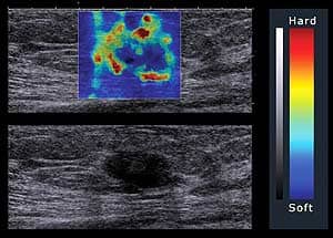
Better Breast Lesion Detection
 |
| Image of a malignant breast lesion, ductal carcinoma in situ, BI-RADS 5, captured by SuperSonic Imagine’s Aixplorer ultrasound system. The Aixplorer is FDA approved and is the only ultrasound system that images with Shear Wave Elastography technology |
Ultrasound has proven its value in breast imaging and has been shown to detect lesions missed by mammography. But ultrasound is not very specific. It finds both benign and malignant lesions, and often biopsy is required to make a diagnosis. Over the years, a variety of ultrasound techniques have been used to improve ultrasound specificity and avoid breast biopsies, including color and power Doppler, harmonics, and compound imaging. Shear Wave Elastography is the newest addition in this continuing development. In general, normal breast tissues are soft and breast cancers are hard. Benign and malignant lesions may look alike on gray scale ultrasound, but they behave differently with elastography. Shear Wave Elastography offers the ability to visualize breast lesions smaller than 5 mm, and it may be possible to differentiate cancer from benign lesions based on tissue elasticity, one goal of an ongoing multicenter international study of more than 2,000 patients. If the study data are supportive, the increased specificity attributable to elastography may help to reduce or even to avoid the expense and risk of unnecessary biopsy. Elastography’s potential to increase the specificity of breast ultrasound is an active area of research for many eminent breast imagers, such as principal investigator Ellen Mendelson, MD, professor of radiology at the Feinberg School of Medicine, Northwestern University. She commented that ?This novel technology enables measurements of tissue stiffness to be obtained in seconds, easily and reproducibly using the same transducer to depict gray scale B mode features of benign and malignant breast masses.?
Monitoring Fibrosis in Liver Cirrhosis
Today, from an imaging standpoint, there is not a sensitive way to monitor the progression of liver fibrosis. MR, CT and conventional ultrasound all lack the ability to detect subtle changes in appearance and behavior of the tissue. These modalities do offer the ability to grossly monitor changes from mild to moderate and severe, but do not allow clinicians to follow patients closely to see the progression of the disease. Monitoring the effectiveness of treatment, whether antiviral, diet, or otherwise, has proven very challenging, and clinicians have tried a variety of methods short of biopsy.
The expectation is that Shear Wave Elastography will be able to, with significant specificity, monitor changes in liver cirrhosis by determining the stiffness of the tissue objectively. This will support better decision-making regarding treatment and provide much needed information on its efficacy. It promises to reduce the number and frequency of invasive procedures such as biopsies with economic patient management and safety implications.
Clinicians are also conducting surveillance of the liver looking for the development of cancer. With conventional ultrasound, a hepatocyte does not appear very dissimilar from hepatocellular carcinomas as the intrinsic contrast is not markedly different. CT and MR do offer contrast agents to aid is this, but the technique still lacks some specificity and is more costly than ultrasound, with risk of radiation exposure over a lifetime using CT if monitoring cirrhosis patients. Shear Wave Elastography promises to provide clinicians with greater diagnostic confidence about whether they are observing a regenerating benign nodule or whether it has more features characteristic of hepatocellular carcinoma. Early detection has pivotal patient management and significant economic implications. This also has serious implications for transplant candidates who have fewer options as tumors grow larger.
Greater Specificity in Prostate Scanning
As ultrasound is used as a primary tool for guiding prostate biopsies, the ability to recognize cellular pathology with great specificity is of paramount importance. Shear Wave Elastography promises to emerge as an important tool in providing better, more accurate biopsies and better yield. Often one can’t distinguish cancer from the background of BPH. Shear Wave Elastography offers the prospect of providing identification of stiffness in areas of the prostate cancer and more efficiently and accurately guiding biopsy. Currently, samples are taken from quadrants and by randomly sampling to capture enough material to attempt to identify cancer. This lack of more focused targeting can result in a missed diagnosis as well as repeat visits and procedures at great time and expense. It is particularly problematic among those with chronic prostatitis who often have elevated PSA values and require multiple visits. Shear Wave Elastography allows clinicians the ability to zero in on specific areas of the prostate for more efficient and effective procedures.
Early Diagnosis and Monitoring Tendons
Today clinicians don’t have effective diagnostic and monitoring imaging tools for tendons. MR is a very expensive way to observe if a tendon is torn, and leading up to that catastrophic event, the modality returns very little information about these structures such as how they are reacting to stress and fatigue and progressively degenerating. Moreover, MR is not sufficiently sensitive in early stages of tendon pathology. With Achilles tendons in particular, patients have few symptoms and pain leading up to tears and ruptures, and by the time MR is performed, they are at a late stage in the condition with greater fallout. There is a need for earlier detection as tendon pathology may progress without symptoms. With millions of patients with tendonopathy, the economic implications are enormous. Patients with progressive tendonopathy could avoid degenerative tendon conditions if clinicians could more easily diagnose and follow them and intervene before the point of tears rupture. Thereafter, treatment becomes more challenging and costly as rehabilitation is long, healing is slow as generally tendons don’t heal well, and surgical outcomes tend to be poor.
Tears tend to occur in specific areas where, for instance, there is hypoxemia, and these are not uniform with micro tears throughout. Scanning these areas in real time with the capabilities of elastography, an entire tendon can be better monitored and changes clearly identified. Interventions such as physical therapy, steroids, and load reduction can be monitored to determine whether the interventions are addressing the stiffness and to what degree. Such monitoring can play a critical role in guiding therapy and preventing a much more traumatic and costly disease progression and treatment spiral. Monitoring to date has been largely subjective, with clinicians depending on patient assessment of how they feel, which is a poor guide and can lead to progressive degeneration as patients don’t always experience symptoms and pain until the tendons are badly damaged. Shear Wave Elastography for the first time offers an objective assessment of stiffness in real time and a better view of the physiology in motion.
Conclusion
As ultrasound advances have, over several decades, provided clinicians with greater tissue imaging specificity, the clinical utility and economic value of the modality have increased. With this new leap forward, Shear Wave Elastography builds upon these advances, offering the next level of tissue specificity. As systems become more available, in the next several years, new horizons of diagnosis will open where clinicians will be able to determine not only the appearance of a pathology but also its actual behavior.
Alda Cossi, MD, is director of ultrasound at Boston Medical Center and associate professor of radiology at Boston University School of Medicine.
Reference
- The quantification tool is available outside of the United States.