Ocentra GYN from Nucletron
Creating a newly developed treatment planning system for brachytherapy, the Ocentra GYN from Netherlands-based Nucletron, in conjunction with the General Hospital Vienna (AKH)/Medical University of Vienna (MUW), is designed for combined intracavitary and interstitial treatment. Using dynamic image-guided brachytherapy, the Ocentra GYN treats patients with locally advanced cervical cancer. It is fully tuned to the GEC ESTRO GYN recommendations via predefined optimization and evaluation settings, and its software contains an applicator library of gynecologic applicators for fast application modulation. With the applicator in place, an imported 3D data set can allow users to calculate the exact needle positions for the optimal plan.
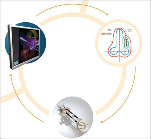
During the treatment, users can visualize the dose distribution and adjust the needle positions. Once the applicator is placed and imaged in situ, a conventional, or “base,” plan is generated using interstitial needles, comparable to conventional postplanning that presets the geometric relation of the applicator to the target. This step is automatically based on an advanced optimization algorithm, Hybrid Inverse Planning Optimization.
 |
The Visible Difference
According to Nucletron, the Ocentra GYN represents the radical new standard in dynamic, image-guided brachytherapy for intracavitary and interstitial applications. The outer surface and the source path are predefined, facilitating the positioning of the applicator by marking points, translations, and rotations directly in the MRI or CT data set. The modeling is so accurate that a 0.0-mm margin around the PTV can be explored while keeping the uncertainty level below 5%.
A drastic reduction of planning time is accomplished through the implementation of the applicator library, which enables users to precisely delineate the patient anatomy and modeling of the applicator. Contour sets can be imported to subsequent fractions by taking the applicator as the reference system. Users can copy target structures that are visible only on MRI onto subsequent CT-based implants if MRI availability is limited. Physicians can study in detail the changes in anatomy of organs at risk in-between fractions and implantations.
The Spec Sheet
- Standardization through Paris system and the Manchestor method
- Conversion to dose-volume optimization using 3D images
- Upfront implant geometry
- Optimized applicator/needle position based on real-time dose guidance
- Interfraction adaptation to patient anatomy changes
- GEC ESTRO recommendations for gynecologic brachytherapy
- Predefined GEC ESTRO DVH parameters and prompt DVH-based analysis


