· Latest Carestream Health PACS Debuts Native 3D Capabilities
· Software Promotes Positive Image of Patient Experience
· Centralized PACS Links Large Health Care System
· Candelis ImageGrid Expands Web-based Offerings
Latest Carestream Health PACS Debuts Native 3D Capabilities
Native 3D imaging and advanced cardiac features will be included in Carestream Health’s latest KODAK CARESTREAM PACS, which offers optional integrated applications such as image fusion and orthopedic surgical templating.
Health personnel can choose to equip their facilities with native real-time 2D and 3D capabilities, including maximum intensity projection (MIP), minimum intensity projection (MinIP), multiplanar reconstruction (MPR), volume rendering, tissue definition, and vessel tracking functionality from a single virtual desktop.
In terms of specialized cardiac functions, users can benefit from cardiac review and analysis, cardiac cage removal in 3D imaging, and cardiac calcium scoring—all tools that can assist radiologists in the analysis of cardiac CT imaging exams. In addition, the PACS permits bone/cage removal to enhance viewing of other structures and vessel analysis enhancements, including extended stenosis marking. It also can come with a native single media archive that includes shelf management support, which the company said will benefit small sites that desire a low-cost solution for data backup and archiving.
According to Carestream Health, the built-in dynamic streaming capabilities of its PACS allow advanced functionality to be readily available to authorized users inside and outside the institution. In particular, clinicians will have the opportunity to review the first images in an exam, while the rest of the study is being transmitted. With the release of a new licensing structure, facilities will be able to purchase the optional features according to the number of concurrent users, rather than have the tools restricted to specific workstations.
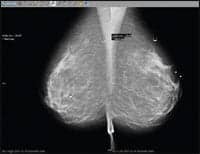
Other options for customers come equipped with native features for the KODAK CARESTREAM Mammography Workstation, which is capable of displaying images from all modalities. These features include an image map, automatic chest wall justification, enhanced annotation capabilities,, and the integration of Confirma CADstream software for breast MRI exams and Cedera B-CAD software for breast ultrasound exams. The enhanced ORTHOVIEW 4.0 orthopedic digital planning solution has support for specialized surgical templates for trauma cases, and an integrated FUSION7D Software from Siemens Medical Solutions delivers enhanced imaging of cancerous tumors and lesions so that oncologists can better visualize anatomic and metabolic information.
Other options for customers come equipped with native features for the KODAK CARESTREAM Mammography Workstation, which is capable of displaying images from all modalities. These features include an image map, automatic chest wall justification, enhanced annotation capabilities,, and the integration of Confirma CADstream software for breast MRI exams and Cedera B-CAD software for breast ultrasound exams. The enhanced ORTHOVIEW 4.0 orthopedic digital planning solution has support for specialized surgical templates for trauma cases, and an integrated FUSION7D Software from Siemens Medical Solutions delivers enhanced imaging of cancerous tumors and lesions so that oncologists can better visualize anatomic and metabolic information.
Software Promotes Positive Image of Patient Experience
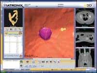
Since acquiring the V3D-Colon 3D imaging software, developed by New York-based Viatronix Inc, Washington Radiology Associates of Washington, DC has performed more than 1,200 virtual colonoscopy (VC) studies throughout Maryland, Virginia, and the District of Columbia.
“The Viatronix software provides highly sophisticated and user-friendly features for the interpretation of VC studies,” said Mark E. Klein, MD, one of the 24 radiologists who work at the practice. “The 3D visualization software is excellent and creates superb images. It allows for comprehensive review of the colon and identification of polyps.”
 |
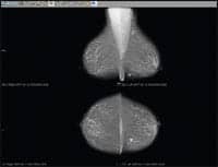
| Viatronix recently introduced upgrades to its V3D-Colon 3D imaging software, including the ability to view the dissected colon segment without distortion or flattening. |
Data has shown that for more than three quarters of America’s population, the sheer thought of a colonoscopy—and the discomfort and inconvenience associated with it—was enough to prevent a person from getting screened for colorectal cancer. Yet, this type of cancer is the second leading cause of cancer death in the United States.
As an alternative to traditional colonoscopy, virtual colonoscopy entails the colon being inflated with carbon dioxide introduced through a small rectal tube. Through this technique, in addition to enhancements to its software technology, Viatronix pledges that the negative image of colonoscopy will be changed forever.
The company’s V3D-Colon 3D imaging software demonstrated a number of upgrades that were unveiled at the annual Radiological Society of North America meeting last November. Health care providers will have the ability to load any two colon image series for review and comparison, as well as view the dissected colon segment without distortion or flattening. Users can edit new “translucent view” color schemes, and reports that are compliant with C-RADS guidelines can be created. Other features include an intuitive user interface, automatic segmentation and centerline extraction, electronic bowel cleansing, automatic and interactive navigation, and 100% lumen coverage and verification.
“Their unique translucency feature allows for rapid differentiation of tagged stool from polyps,” Klein said. “The software also monitors what areas have been reviewed and creates a list of missed regions, which can then be simply and rapidly evaluated by the radiologist.”
The technology is also cost-effective, according to the company, who points out that the new procedure can bring about increased revenue, faster return on investment, and the increased utilization of CT.
“We deliver high-end organ-specific clinical applications developed around a physician’s workflow,” said Zaffar Hayat, president of Viatronix Inc. “In the near future, we expect to deliver additional organ-specific modules to meet the demands of the radiology market.”
Centralized PACS Links Large Health Care System
Set to be released this spring, ProSolv version 4.0 from Fujifilm company ProSolv Cardiovascular, Indianapolis, aims to bring about critical Web-based access to all the features and functionality of the PACS, complete with security protocols, remote reading capabilities, and pediatric Z-scores.
This up-and-coming development stems from the success of the company’s ProSolv Cardiovascular, which recently was ranked number one by KLAS in the 2007 Cardiology PACS study. Earning top accolades in the majority of 40 performance indicators, ProSolv was touted for its commitment to technology, ease of implementation and support, third-party vendor compatibility, and timely enhancement releases.
“The ProSolv application is unique because it is truly a Web-based system,” said Karen Bower, a marketing/product manager at the company. “ProSolv also excels with our unrivaled reporting capabilities.”
With the ProSolv CardioVascular cardiovascular image and information system (CVIIS), users can enjoy seamless integration of every modality, and are able to review patient studies and create comprehensive, customizable reports from a single workstation. Due to its cascadable architecture, the offering is also scalable, being able to expand from a single server to large multiple server networks as needs arise.
Bower said because the application is completely Web-based, the company has been able to quickly react to the demands of the market. Customers can enjoy the latest technologies through Web-based upgrades while retaining historical data. “We have had the opportunity to move fluidly with the rapidly evolving market, bringing changes to our application as a result of customer feedback, much more swiftly than a lot of our competitors,” Bower said.
Also included are reporting functions that auto-summarize, auto-fill, and auto-interpret, among other capabilities. Tools allow for wall-motion scoring, coronary tree diagrams, and freehand drawing. With the neutrality of vendor and hardware, with robust interfaces to the EMR, HIS, and CPOE, final results are immediately available, Bower pointed out.
“Even in the realm of cardiology, an interventionalist and an echocardiographer have different needs, different ways of reporting, most specifically what clinical tools are needed, what systems need to be interfaced to,” Bower said, adding that with the release of Version 4.0 and Fujifilm’s Synapse PACS integration, radiologists can seamlessly access all cardiology images and information while working within Synapse. Meanwhile, cardiologists are able to access all Synapse radiology data—all from one workstation with a single sign-on.
“Versatility is what each modality needs to drive the road map, what partners we work with,” Bower said. “The product’s design continues to be driven by the ?unknown’ in the market, as to exactly what a CVIS is and should be. We need to continue, and have proven to do this well, to be quick on our feet, as we determine the long-term road map.”
Candelis ImageGrid Expands Web-based Offerings
Medical informatics company Candelis Inc, Irvine, Calif, recently introduced a new suite of RIS/PACS technology capabilities with general radiology and mammography-specific applications for its ImageGrid DICOM appliance.
 |
| MLO/CC Standard Hanging |
The ImageGrid PACS Web Viewer and ImageGrid PACS Mammography Web Viewer were both developed to streamline diagnostic study reading. Promoting easy access for its users, the PACS viewer offers a variety of query/retrieve capabilities, which include multiple, side-by-side study viewing, fast retrieval of multigigabyte studies, accelerated streaming of multiframe studies, customizable prefetching of relevant priors, and advanced layout management. With the ImageGrid PACS Mammography Viewer, physicians can make use of customizable mammography hanging protocols, user-defined mammography report flow, and integration with mammography CAD.
“We have rapidly seen that the appliance architecture for RIS/PACS is a key requirement for customers who are either looking at a first-time implementation or looking to replace an existing RIS/PACS,” said Mazi Razmjoo, Candelis vice president of sales. “Candelis’ industry-first appliance architecture has the benefit of providing a cost-effective, fully integrated, and simple to install and operate solution with a significantly lower total cost of ownership.”
 |
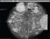
| MLO plus CAD markings for iCAD |
According to Candelis, the complete end-to-end digital image management appliance solution meets the needs of budget-conscious hospitals and imaging centers. One such example is the Virginia Mason Medical Center, based in Seattle. The facility already possessed a separate PACS, albeit one that did not sufficiently support the center’s mammography efforts. When the computer staff sent out initial queries to vendors it was considering to provide them with a tailored solution, Candelis’ responsiveness stood out right off the bat.
“They were very flexible,” said Courtney Allen, VMMC PACS administrator. “They made modifications on their end to match up on what they needed to do to make messages come through.”
Allen said he was particularly seeking the ability to presearch from VMMC’s current PACS and filter out stored processing raw data images without generating errors. Additionally, he was looking for a modality worklist solution that would help fix one of the center’s workflow problems. Allen said he found that Candelis’ ImageGrid application offered powerful logic functions that enabled him to build intelligent prefetching and routing protocols, known as “rules.”
 |
| CC Hanging plus R2 CAD magnifier |
After VMMC completed its conversion to Candelis’ technology, its digital mammography capabilities expanded by 100%, Allen said. Now, users can preprogram a variety of scenarios, such as load-leveling and special scheduling, and activate them as necessary. For example, Allen said he can enter a prefetching rule to read incoming order messages from the RIS and store a PACS query-retrieve request, which is set for a future date and time. Studies that meet the request’s criteria are then retrieved from the PACS and sent to one or more workstations, depending on the scheduled study location. Portions of a study can be suppressed from a certain destination if need be, and separate rules dealing with acquisition can route new study data coming in to multiple locations. It relieves the workstations and 225 or so modalities that VMMC operates from additional overhead.
“The great part is all of this can be done from practically anywhere with a really slick, Web-based graphical user interface that doesn’t require me to be a programmer,” Allen said. “You just have to have a good grasp of the departmental workflow. There is a full complement of useful tools and utilities for testing, monitoring, configuring, and doing ad-hoc a-b-c retrieval and routing between nodes.”
Candelis also released a new Web-based RIS solution, which integrates with the ImageGrid PAS appliance, and it debuted an ImageGrid DICOM Encapsulation feature, which facilitates non-DICOM file support for paperless imaging.
 |
| The ImageGrid PACS Mammography Web Viewer was developed to streamline diagnostic study reading. |


