 |
· ARIA BIS System Adds Biopsy Capability
· Sonography Effective in Diagnosis of Carpal Tunnel Syndrome
· Ultrasound System Takes Trip Around the World
· Breakthrough Technology Combines ICE with 3D Mapping
ARIA BIS System Adds Biopsy Capability
Advanced Imaging Technologies, Richland, Wash, recently announced that its ARIA Beast Imaging System (BIS), a real-time, through-wave ultrasound imaging system, now comes equipped for image-guided biopsy. ARIA BIS effectively penetrates dense tissue, is automated with whole-breast presentation capabilities, and offers high spatial and contrast resolution.
“If you compare our system to traditional breast ultrasound, they’re using the reflective component of the wave,” explained Michael Hartwig, COO of Advanced Imaging Technologies. “We use the wave that passes through the anatomy. We measure where it absorbs and defracts and combine that with holography to create our image, and we do that at around 100 holograms per second, and capture the images at 15 frames per second.”
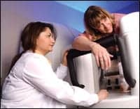 |
| ARIA BIS features real-time image-guidance capabilities that provide physicians with precision for greater biopsy accuracy. |
ARIA ultrasound is FDA-cleared for pediatrics, orthopedics, vascular imaging, and image guidance, but it’s especially valuable in breast imaging, where dense tissue has always been an obstacle. “Our technology has been proven to have superior spatial and contrast resolution, especially in dense tissue,” said Jeanette Griscavage-Ennis, PhD, manager of clinical applications at AIT. “The data we capture is saturated with critical information that is important for diagnostic procedures such as biopsy. Our real-time image-guidance capabilities provide physicians with precision for increased biopsy accuracy.”
So why is ARIA BIS superior to other modalities? Because it combines the best attributes of other technologies, according to Hartwig. “What’s unique is we can do large-area imaging as well as focused imaging,” he said. “MR and CT are nice large-area modalities, but they can’t focus. Ultrasound does focused imaging, but not large imaging. We do both. We also provide information on anatomical structures that’s not available in other images.”
And with so much research documenting mammography’s inability to detect lesions within dense tissue, that’s an important advantage. “The breast is very complex to image because of the large amount of structure within the anatomy,” Hartwig said. “Where other modalities have a difficult time imaging that, we do it very well. The more complexity, the more structure, the better.”
Additionally, the ARIA BIS allows for retrieval of priors for quick cross-correlation with known landmarks, enabling better radiologist workflow. “We allow the radiologist to stay in the reading room instead of getting up in the middle of the ultrasound,” Hartwig said. “We automate the process and provide standardized image sets.”
AIT won its biopsy clearance from the FDA in 2002 and introduced the capability at last year’s annual RSNA meeting. “We provide the opportunity to do high-risk women, high-risk screening, high-risk detection, or biopsy, all in one,” Hartwig said. “And the thing we have going for us as well is that our exam times are very comparable to a mammogram.”
For now, reflective ultrasound is a popular adjunctive diagnostic modality in breast imaging. But the results of ACRIN-6666, which looks at screening breast ultrasound in the high-risk population, suggest the modality could be used for screening in dense tissue imaging in the near future, Hartwig said.
“Our sensitivity in the studies we’ve done in dense tissue is in the 95% range,” Hartwig said. “We’ve outperformed mammography in both sensitivity and specificity.”
—Cat Vasko
Sonography Effective in Diagnosis of Carpal Tunnel Syndrome
The diagnosis of carpal tunnel syndrome when accomplished through sonography is similar in accuracy to electromyographic testing, and it is preferred by more patients, according to a published report in the January issue of the Journal of Neurology, Neurosurgery, and Psychiatry.
 |
Still, Leo H. Visser, MD, PhD, cautioned to Reuters Health, “the value of sonographic follow-up after surgery requires prospective examination.”
Visser, who works for the Department of Neurology at St Elisabeth Hospital of Tilburg, the Netherlands, and his associates prospectively studied the diagnostic value of sonography and EMG in 168 patients with carpal tunnel syndrome, 25 patients with musculoskeletal pain, and a control group of 137 volunteers.
The report states that the cross-sectional area of the median nerve at the carpal tunnel inlet, rather than the distal one third of the forearm, was significantly increased in the patient group, as measured by sonography.
Specifically, sonography was 78% sensitive and 91% specific in diagnosing carpal tunnel syndrome, according to the report. Depending upon the parameters, the sensitivity of EMG ranged from 54% to 82% and its sensitivity ranged from 88% to 91%.
The authors also noted that adding EMG results did not significantly increase diagnostic accuracy of sonography.
Furthermore, more patients said they favored sonography over EMG, according to the researchers. “Sonography is probably preferable because it is painless, cheap, easy to apply, easily accessible,” the report concludes.
In another study, whose findings will soon be published, Visser and fellow researchers sought “to assess (1) the change in the size of the median nerve at the proximal carpal tunnel after surgery compared to after conservative treatment and (2) the correlation between sonographic characteristics and clinical outcome after surgery.”
—Elaine Sanchez
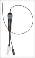
Ultrasound System Takes Trip Around the World
Az.one ultrasound system that was recently shipped will not reach its ultimate destination for another 114 days. But manufacturer ZONARE Medical Systems is perfectly OK with that estimated time of arrival. The unit is traveling around the world, after all.
 |
| ZONARE?s z.one ultrasound system is being used for a 4-month study on board the USS Amsterdam. |
On board the USS Amsterdam, a flagship of the Holland America Line, the equipment embarked on a 114-day cruise around the world, setting sail this past January from Fort Lauderdale, Fla. It will be used during a 4-month study, whose outcome may determine the future use of ultrasound imaging on passenger cruises.
“When medical emergencies arise during a cruise of this length, sometimes we must decide if we need to divert from our set itinerary to the closest port offering appropriate medical facilities,” said Carter Hill, MD, medical director of Holland America. “We want to do what is best for the patient. With advanced diagnostic technology available, such as the z.one system and trained providers, we should be able to make more informed decisions of these kinds as to whether we can treat a patient aboard and a diversion is unnecessary.”
Hill said he selected the z.one system for the study because of its flexible nature, made possible by its convertible ultrasound platform. Users can convert the z.one system at the touch of button from a full-featured, cart-based system into a premium compact ultrasound system. The product is designed to allow for versatility in a variety of clinical settings without sacrificing image quality.
“If a patient feels too ill to come to the ship’s infirmary, the z.one system can easily be taken to the state room, and the exam [can be] conducted there,” Hill said. “As future advances occur, we can also upgrade the system to provide the latest clinical applications and continue to offer state-of-the-art ultrasound imaging for our passengers. This is a tremendous economical and clinical benefit.”
According to ZONARE, the z.one system’s Zone Sonography software-based architecture enables continual improvement as greater processing power becomes available. System upgrades can be downloaded through the Internet.
An alumnus of the Universities of Minnesota and Washington, Hill has been practicing emergency medicine for 28 years and has led Holland America’s medical operations for 2 decades.
The cruise will make its journey through the Panama Canal, Tahiti, Australia, Asia, the Black Sea, and Europe before heading home to Fort Lauderdale. Periodic updates of its travels will be posted on ZONARE’s Web site, www.zonare.com.
—C. Vasko
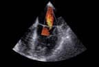
Breakthrough Technology Combines ICE with 3D Mapping
Working in Southlake Regional Health Center, Ontario, Yaariv Khaykin, MD, and Atul Verma, MD, must handle a yearly average of 600 ablations at their busy electrophysiology center. Therefore, for them, intracardiac echocardiography (ICE)—which delivers an ultrasound beam from a linear array of crystals at the tip of a navigable catheter—has been indispensable, allowing physicians to tackle a high volume of complex cases such as atrial fibrillation and ventricular tachycardia ablations.
 |
| New tools to aid in diagnosis and treatment of cardiac arrhythmias. |
However, a new product from Biosense Webster Inc, Diamond Bar, Calif, enabled them to go one step further, integrating real-time ultrasound with 3D imaging. Last year, the Southlake Regional Health Center became the first site in the world to use the company’s CARTOSOUND Image Integration module and SOUNDSTAR 3D Catheter in clinical care. Now, electrophysiologists everywhere can use the technology with the new Carto XP Navigation System to aid them in the diagnosis and treatment of cardiac arrhythmias.
“While ICE allows you to see cardiac and extracardiac structures, such as the esophagus, in real time, it does not allow tagging sites of interest, anatomical landmarks, or sites where ablation lesions have been placed,” said Khaykin, who, along with Verma, trained at the Cleveland Clinic under the tutelage of ICE pioneer Andrea Natale, MD. “CARTOSOUND integrates ICE with 3D imaging, allowing for registration of 2D ultrasound slices in 3D space. This allows us to rebuild real-time anatomy, sort of like putting an apple back together from slices. Once the anatomy is built, we can instrument the chamber of interest with a catheter and manipulate the catheter without using fluoroscopy.”
Although conventional 3D imaging permits 3D virtual reconstruction of mapped structures, Khaykin explained it requires point-by-point mapping. This task, he continued, is a “time-consuming procedure, further complicated by the fact that moving the catheter from point to point actually distorts the anatomy and integration of preacquired CT and MRI imaging.” Likewise, speeding up the process may be inaccurate because images are frequently not up to date.
 |
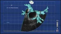
The new CARTOSOUND Module integrates real-time ICE images from the Siemens ACUSON Sequoia and Cypress ultrasound systems using the SOUNDSTAR 3D Catheter and CARTO XP EP Navigation System. According to Khaykin, this navigation system uses GPS-like technology to locate catheters in space, usually placed inside the patient. Additionally, three magnets that generate orthogonal magnetic fields are placed under the patient. Each catheter is equipped with one or more navigation sensors, comprised of three orthogonal coils. The system uses these coils to triangulate the location of each catheter in space.
A similar navigation module was placed at the tip of the ICE catheter, so that each acquired 2D image has 3D coordinates, Khaykin continued. After contours of the structure are drawn on the 2D image, they are displayed in 3D space and combined to form a 3D image.
The technology has the capacity to become the future of cardiac arrhythmia treatment, as physicians endeavor to treat more patients with progressively more complex arrhythmias. “These procedures carry many inherent risks, and success is not a given,” Khaykin said. “Any tools that can help perform these procedures faster, better, with a lower risk and less fluoroscopy exposure will be met with enthusiasm by the medical community.”
—E. Sanchez
