
 |
By Elaine Sanchez
· Shaving Seconds Saves Lives
· ACR Responds to Report: CT Study Stirs Up Controversy
· Study Alert: Worst Curve Algorithm Helps Determine Malignancy
Shaving Seconds Saves Lives
In emergency situations, time is crucial, and those who work in a hospital’s intensive care unit know firsthand that minutes, or even seconds, cannot be spared. So instead of spending crucial time transporting patients to a particular exam room, why not bring the equipment to them?
Upon pondering this question, The Moses H. Cone Memorial Hospital, of Greensboro, NC, arrived at a solution that 49 other hospitals around the world have turned to—the CereTom portable CT scanner, from NeuroLogica Corp of Danvers, Mass.
 |
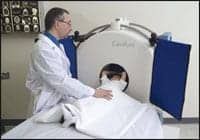 |
| With the CereTom portable CT scanner, doctors are able to get high- quality CT images without being forced to move patients to the traditional hospital CT scanner. |
“Having access to portable CT scanning in our neurosurgical ICU enables us to bring diagnostic imaging technology directly to the bedside for this patient population,” said Steve Shanaberger, director of radiology at The Moses H. Cone Memorial Hospital. “The CereTom allows the nurse to remain on the unit and improves care by eliminating the need to coordinate the transportation of critically ill patients to the fixed scanner in the department.”
As the 50th hospital to purchase the CereTom, The Moses H. Cone Memorial Hospital will have access to a compact, lightweight piece of equipment that enables physicians to gain critical diagnostic imaging of patients immediately following surgery. These doctors will be able to obtain high-quality CT images without being forced to move patients to the traditional hospital CT scanner.
The eight-slice CT scanner is not just limited to the ICU. Rather, it can be used in the radiology department, emergency department (ED), operating room (OR), NICU, MICU, SICU, interventional suite, or any medical clinic. Portable and lightweight, the CereTom is 29 inches deep, 5 feet tall, and 4 feet wide.
Currently, hospitals use the CereTom to diagnose head trauma and stroke patients immediately upon arrival to EDs, and it can also help diagnose, monitor, and potentially fix any complications prior to transporting a patient outside the OR. Additionally, with the CereTom, pediatricians can monitor the health of babies in the neonatal and pediatric ICUs. Two National Football League teams, the Oakland Raiders and the Indianapolis Colts, used a CereTom unit during the 2007-08 season.
“Announcing our 50th customer demonstrates the growing demand for this unique imaging technology,” said Eric M. Bailey, NeuroLogica president and CEO. “This milestone validates the importance of bringing the diagnostic capabilities of CT scanning directly to the patient.”
Acknowledging another major feat, NeuroLogica recently revealed that the CereTom was employed in Saudi Arabia during a successful separation surgery of 1-year-old twins who were conjoined at the skull. Through the CereTom, a surgical team successfully checked for bleeding and hydrocephalus, and monitored the condition of the twins’ spinal fluid and swelling.
“Having the portable CT scanner in the OR allowed for on-the-spot evaluation and eliminated the need to transport the twins to radiology following the surgery,” said Ahmed Al Ferayan, MD, the neurosurgeon who performed the surgery. “Instead, we were able to take them directly to the ICU for monitoring and recovery.”
ACR Responds to Report: CT Study Stirs Up Controversy
Amid the hustle and bustle of the week-long RSNA meeting, an article was published in the New England Journal of Medicine, shining a light on potential cancer risks associated with CT scans.
 |
Not only did it create a buzz in the medical imaging community, but a mass media frenzy followed, with the issue getting play in the country’s major newspapers as well as on influential TV news broadcasts.
Picking up the story, the Associated Press wrote, “Millions of Americans, especially children, are needlessly getting dangerous radiation from ‘super x-rays’ that raise the risk of cancer and are increasingly used to diagnose medical problems, a new report warns.”
Addressing the general public’s heightened alarm, the American College of Radiology issued an official response to topics raised in the November 29 study, titled “Computed Tomography—An Increasing Source of Radiation Exposure.”
“The American College of Radiology is concerned that certain conclusions and comparisons made in the study ? may be inappropriate and cause patients to mistakenly avoid getting life-saving medical imaging care,” the written statement asserted.
In the report, authors David Brenner and Eric Hall of Columbia University Medical Center in New York wrote that doctors, underestimating the radiation risk from CT scans, may be ordering an excessive amount of them. In fact, up to one-third of the scans may be unnecessary, they concluded.
“If it is true that about one-third of all CT scans are not justified by medical need, and it appears to be likely, perhaps 20 million adults and, crucially, more than 1 million children per year in the United States are being irradiated unnecessarily,” according to the published study. It also states that as many as 2 percent of all cancers in the United States may be caused by radiation received from CT scans, but the authors point out that there are no studies directly linking CT scans to cancer. Brenner and Hall offer ultrasound and MRI as safer options that doctors can employ.
In its response, the ACR was particularly concerned with a comparison drawn in the study, equating radiation exposure experienced by atomic bomb survivors in Japan to patients receiving CT scans today.
“You cannot equate the Hiroshima experience to a CT scan over a period of time,” said Arl Van Moore Jr., ACR chairman. The College outlined key differences between the two populations, such as the type of radiation exposure each experienced. CT exams expose patients solely to x-rays, the College wrote, while atomic bomb survivors were exposed to x-rays, particulate radiations, neutrons and other radioactive materials.
Ultimately, the ACR contended, patients may be confused or distressed by statements in the study.
“For example, the ACR has long opposed full-body CT scans for asymptomatic patients, one of the exams that the NEJM article authors put forth as a driver of future CT growth,” Moore said. “We also support the ‘as low as reasonably achievable’ concept, which urges providers to use the minimum level of radiation needed in such exams to achieve the necessary results.”
However, Moore also points out that there are upsides to the published study and the controversy it has generated. It raises awareness on a public health issue, Moore said. Patients could become educated on talking with their physicians about CT’s risks, benefits and possible alternatives, and finding out if their exam is being done in an ACR-credited facility.
The ACR urged both patients and providers to visit its official Web site’s “Radiology Safety” section, which it co-manages with the Radiological Society of North America.
Study Alert: Worst Curve Algorithm Helps Determine Malignancy
A study evaluating kinetics of suspicious breast MRI lesions has suggested that the patented “worst curve” algorithm of the CADstream system, developed by Confirma, may serve a role in determining malignancy.
 |
Researchers at the Seattle Cancer Care Alliance and the University of Washington worked together, under Lilian Wang, MD, to compare three different computer-aided evaluation kinetic features of the suspicious lesions. The group then determined the feature that best predicts benign and malignant outcomes.
According to findings, the most suspicious curve as identified by CADstream was significantly different between benign and malignant lesions. The study also discovered that any washout enhancement was associated with malignancy in nearly half of the lesions.
“Our findings are consistent with the ACR BI-RADS Atlas recommendation for reporting the ‘worst looking kinetic curve’ of a lesion with breast MRI,” said Wang, a radiology resident at the Seattle Cancer Care Alliance and University of Washington.
In the study, called “MRI Detected Suspicious Breast Findings: Comparison of Kinetic Features Measured by Computer-aided Evaluation in Benign and Malignant Lesions,” Wang and her team retrospectively examined 125 suspicious lesions for which computer-aided evaluation via CADstream and subsequent image-guided biopsy had been performed. These lesions, 42 of which were malignant and 83 benign, were selected out of a pool of 1,538 breast MRI examinations conducted between November 2004 and November 2006.
Other researchers who worked on this study include Wendy DeMartini, MD, Constance Lehman, MD, and Sue Peacock, MSc.
In the July 2007 issue of Radiology, joint research from the University of Washington and Seattle Cancer Care Alliance was published, indicating that CADstream for breast MRI may improve standardization and analysis of benign and malignant lesions.
Results of the study focusing on kinetics were recently presented at the annual meeting of the Radiological Society of North America in Chicago.
 |
At the annual meeting of the North American Society of Cardiovascular Imaging (NASCI), Raleigh, NC, held in October in Washington, DC, radiologists from the University of Cincinnati presented a new protocol for thoracic imaging: contrast-enhanced MDCT. Christopher Meyer, MD, and Achala Vagal, MD, discovered the technique for more precise imaging of chest vasculature.
“We found that the rapid-imaging scanners were almost too fast for venous studies,” said Vagal, a University of Cincinnati assistant professor and radiologist at University Hospital. “By the time the contrast reached the patient’s veins, there were too many artifacts to make any meaningful conclusions about possible disease—for example, blood clots.”
The protocol developed by Vagal and Meyer addresses this issue by using two different doses of contrast. The first dose is 140 cc of undiluted contrast, and the second contains a diluted mixture, 100 cc of contrast mixed with 10 cc of saline solution.
“The key to getting accurate clinical images of the veins is in the timing,” Vagal noted. So the second syringe of contrast is administered immediately following the first, both at a rate of 4 cc per second. Then 60 seconds are left between the final contrast injection and beginning the CT scan.
“Previously, there was so much dense contrast in the veins that all you could see on the CT scan were streaks that didn’t tell you anything about possible venous disease,” said Vagal. “Delaying the scan gave us enough time for both the arteries and the veins to be opacified, which resulted in the crisp images that allowed us to make better clinical determinations.”
The role of CT in thoracic imaging has been on the rise in recent years as the modality has offered increasing speed and precision. At the 2006 RSNA meeting, Geoffrey D. Rubin, MD, of the Department of Radiology, Stanford University School of Medicine, Stanford, Calif, presented on 64-slice MDCT for imaging of the chest. He noted that the increased resolution offered by slices as thin as 1 mm, coupled with the power of volumetric reconstruction, have led to extremely enhanced visualization of chest structures.
In addressing vascular imaging in the chest, Rubin noted that gating reveals the physiology of vessels, including dissections and calcifications that were impossible to detect before. Gating enables the visualization of the clots that create acute coronary syndrome.
“Venous disease is rare and can be difficult to pinpoint,” Vagal noted. “This new protocol uses the same imaging equipment in a novel way that allows us to acquire better venous images and make good clinical decisions.”
—Cat Vasko

