|
by Cat Vasko
· IMRT: Fewer Side Effects for Females
· Aurora Welcomes Nobel Laureate to Advisory Board
· Combating Cervical Cancer
· Organon and Philips Team on Study of Drug Effects at the Molecular Level
· The Cutting Edge: Intermountain Implements Gamma Knife Perfexion
· Spotlight on Study: Early Diagnosis, Prevention with MRI
· Research Alert: CardiArc SPECT System Up for Clinical Trials
IMRT: Fewer Side Effects for Females
 |
A study in the August 1 edition of the International Journal of Radiation Oncology*Biology*Physics, the official journal of the American Society for Therapeutic Radiology and Oncology, reports that women with early-stage breast cancer who receive intensity modulated radiation therapy (IMRT) develop significantly fewer side effects than women who receive traditional radiation therapy.
The study, titled “Intensity-Modulated Radiotherapy Results in Significant Decrease in Clinical Toxicities Compared With Conventional Wedge-Based Breast Radiotherapy,” reviews the acute and chronic toxicity of IMRT compared with conventional wedge-based treatment, which can result in side effects like changes to the breast’s shape and feel or skin irritations similar to a sunburn. Although these side effects usually fade when treatment has ended, there is a risk of long-term side effects such as changes in the color, texture, or firmness of the skin or breast swelling.
Physicians at William Beaumont Hospital, Royal Oak, Mich, evaluated 172 women with early stage breast cancer as part of the study. One group (54%) received IMRT, while the control group received conventional wedge-based radiation. The researchers used a static multileaf collimator to deliver the IMRT, in an effort to optimize dose homogeneity and decrease normal tissue toxicity.
Overall, women who received IMRT reported significantly fewer breast-related side effects compared with women who received wedge-based therapy. Less than half of the IMRT group had notable reddened or itchy skin, compared with 85% of the women in the control group. Only 1% of the IMRT group experienced breast swelling, compared with 28% of the other group. Five percent of the IMRT group had changes in skin color, while 50% of the control group had hyperpigmentation. Eight percent of the women in both groups reported pain as a result of the treatment.
“Earlier studies have demonstrated the benefits and importance of radiation therapy in the treatment of many women with breast cancer,” said Asif Harsolia, MD, lead author on the study and a radiation oncologist with the Permanente Medical Group in Santa Clara, Calif.
“It is exciting that we are now conducting studies with the goal of helping to make these treatments easier and more comfortable for women. This study, along with other recent data presented by our colleagues, demonstrates that improving dose homogeneity within the breast with IMRT results in significantly fewer side effects for women undergoing radiation therapy for early stage breast cancer.”
—Cat Vasko
Aurora Welcomes Nobel Laureate to Advisory Board
 |
| Dr. Aaron Ciechanover, MD, Dsc |
On August 13, the 2004 Nobel laureate in Chemistry, Aaron Ciechanover, MD, Dsc, joined the advisory board of Aurora Imaging Technology Inc, North Andover, Mass. Ciechanover is a Distinguished Research Professor of Biochemistry in the Rappaport Faculty of Medicine and Research Institute of the Technion-Israel Institute of Technology, Haifa, Israel; his Nobel-prize-winning research involved the discovery of the ubiquitin system for intracellular protein degradation.
“We don’t believe we’re just a medical technology company,” said Olivia Ho Cheng, president and CEO of Aurora. “Our focus is breast cancer, and we want to improve detection and maybe eventually find its root cause. We jokingly say that we are working to work ourselves out of jobs.”
The ubiquitin system is the process by which the body marks cells for destruction; without this molecular label, a cell could live on indefinitely. “It’s the finding of a needle in a haystack,” Cheng said. “Having this understanding of how our body works, how cells are born and die, really helps the research to find drugs that can kill cancer—drugs that can mark cancer for death.”
Ubiquitin modification of cellular targets is involved in a wide array of cellular processes, including cell cycle and division, apoptosis, transcription, maintenance of cellular quality control, modulation of metabolic pathways, and more. Aberrations in the ubiquitin system underlie the pathogenesis of many diseases.
“Our research focuses on the involvement of the ubiquitin proteolytic system in the regulation of transcriptional activators and apoptosis,” Ciechanover said. “Both subjects are tightly linked to cancer pathogenesis. Transcriptional regulators are involved in both tumor progression and suppression, and their degradation plays a major role in their regulation. Apoptosis is an important process via which malignant cells normally die, and evasion of apoptosis is one of the most basic mechanisms that underlies tumor growth and that is regulated by the ubiquitin system.”
Cheng explains that this research will be a useful contribution to Aurora’s quest to identify compounds that could indicate angiogenesis at an early phase. “We hope that we can find these compounds and be able to detect them through MRI, making detection easier and more precise,” she said. “That’s why we looked for Dr. Ciechanover. We want to be able to identify compounds more closely related to breast cancer.”
And Ciechanover notes that ubiquitin serves as a rich platform for drug development. “Since the ubiquitin system we discovered regulates numerous processes in the cell, it is not surprising that disorders in the system underlie the pathogenesis of many diseases, including numerous malignancies,” he said. “One anticancer drug [Bortezomib] has already been developed and is on the market, and many more either are on the way or will be developed.”
—C. Vasko
Research Alert:
 |
Combating Cervical Cancer
While the new vaccine for cervical cancer caused some controversy among parties concerned with the privacy rights of young women, new developments in cervical screening and detection remain a priority. In the United States—where cervical cancer strikes about 10,000 women a year and causes up to 4,000 deaths annually—the advent of liquid-based cytology in the mid 1990s revolutionized the conventional Pap smear. It provided a screening method considered significantly more effective than the conventional Pap smear in detecting precancerous lesions.
Today, we know that imaging technology has made a difference in detection. In 2003, the FDA cleared the ThinPrep Imaging System from Cytec Corporation to make it easier for cervical cancer lesions and other abnormalities to be detected. With approximately one third of false-negative Pap results being due to abnormal cells that were missed or misclassified, the ThinPrep Imaging System is the first fully integrated, interactive computer system that assists cytotechnologists and pathologists in the primary screening and diagnosis of ThinPrep Pap Test slides. The ThinPrep Imaging System combines imaging technology with human interpretive expertise to produce a screening method that is more effective and efficient so patients can seek treatment before abnormalities develop into cancer.
New research evaluating the ThinPrep Imaging System was featured in a recent issue of the British Medical Journal (BMJ). Coconducted by the University of Sydney and Douglass Hanly Moir Pathology in Sydney, the study reported that “the ThinPrep Imager detected significantly more histological high-grade squamous disease than did manually read conventional cytology slides.” The authors compared the results of split-sample slide pairs from 55,164 patients over an 11-month period. In each case, the conventional smear was prepared first and the ThinPrep Pap Test slide was prepared with the remaining material. The ThinPrep slides were processed using the ThinPrep Imaging System.
“The significant increase in precancerous lesion detection seen with the ThinPrep Imager in this study is very positive for cervical cancer prevention and provides evidence health care professionals in Australia have been waiting for,” said coauthor Annabelle Farnsworth, MD, Douglass Hanly Moir medical director.
In the United States, the ThinPrep Pap Test is currently the only liquid-based cytology method approved by the FDA for human papillomavirus (HPV), Chlamydia, and gonorrhea (CT/NG) testing, and is the most widely used method for cervical cancer screening in the country. The tragedy of cervical cancer is that it often strikes when a woman is still young. Many of these victims may be women who have not yet given birth, and cervical cancer treatment may make future fertility impossible. Early detection—through imaging technology—is saving lives here and abroad.
 |
| Rick Harwig, CTO, Philips |
Organon and Philips Team on Study of Drug Effects at the Molecular Level
Philips Medical Systems’ European counterpart has joined forces with N.V. Organon, Oss, Eindhoven, the Netherlands, to develop new drugs and therapies for mental disorders and cancer. Using biomarkers and Philips’ medical imaging technology, researchers hope to study the effects of psychiatric drugs on the brain at the molecular level.
Using molecular imaging technologies could expedite the development and approval of new drugs and therapies by measuring drug effects at the molecular level. In addition, these imaging technologies can be used to monitor the effect of the therapy and customize the treatment program accordingly. Such customized treatment programs could increase effectiveness and comfort, and ultimately accelerate and improve patient outcomes.
“Organon has invested heavily in optimizing its R&D efforts,” explained David Nicholson, executive vice president research and development, of Organon. “We are convinced that biomarker research will accelerate the R&D process and improve the success rate of developing new molecular and biological therapies. In particular, our drug development programs for the treatment of psychiatric and immune disorders are expected to benefit from this collaborative research effort with Philips.”
“Our medical imaging modalities are rapidly improving and have evolved into extremely powerful tools to image the function and behavior of an anatomical feature, and not just its shape,” said Rick Harwig, chief technical officer (CTO) of Philips. “The combined technological expertise of Philips and the life sciences know-how of Organon will definitely speed up the evolution of our imaging modalities into tools to image the body at the molecular level. In addition, they will enable new opportunities in molecular diagnostics.”
As part of the agreement, scientists from Organon will work at the Philips Life Sciences Facilities, Eindhoven, to identify, validate, and exploit novel biomarkers.
The Life Sciences Facilities is a multidisciplinary research facility, which provides the necessary biological, chemical, and technical infrastructure and expertise for research and development in the field of translational biomarkers and molecular medicine.
The Cutting Edge:
Intermountain Implements Gamma Knife Perfexion
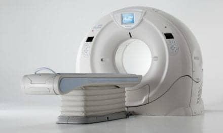
On August 14, the Jon and Karen Huntsman Cancer Center at Intermountain Medical Center, Murray, Utah, saw the first use of its latest radiation treatment technology purchase—the Leksell Gamma Knife Perfexion from Elekta, Norcross, Ga. Stereotactic radiosurgery, including the procedures performed using the Gamma Knife, is a noninvasive method for treating brain disorders. The extreme precision of the Gamma Knife makes it possible to administer a high radiation dose to the diseased area, minimizing the risk of damaging healthy tissue.
 |
| The Leksell Gamma Knife Perfexion is being used at The Jon and Karen Huntsman Cancer Center. |
“The Gamma Knife is a method of delivering a very accurate, very high dose of radiation to targets in the brain, using high-res imaging from MRIs and CT scans,” said Gordon Watson, MD, PhD, director of the radiosurgery program at Intermountain. “We can target any point in the brain with this highly focused radiation and give a high enough dose that whatever we’ve targeted is killed. It allows us to remove a tumor without having any surgery.”
Watson explains that the Gamma Knife can replace surgery only for specific indications—tumors must be localized, less than 4 cm in size, and sufficiently distant from critical structures. “Even though the dose is very precise, there’s still fallout,” he noted.
But the device is user-friendly and intuitive, Watson said: “After going through a very intense weeklong training course, everybody’s feeling quite comfortable with it.” In its first 2 weeks, the Gamma Knife was used to treat eight patients. Intermountain hopes to treat 100 patients this year, and 200 patients a year after that. “This is a very cost-effective way to treat these patients,” Watson noted.
It is also time-effective. Open brain surgery could take anywhere between 1 and 8 hours in the OR, followed by 2 to 5 days of recovery in the hospital. Gamma Knife treatment is quite different.
“If a patient comes into the hospital at 7 am for the gamma knife, they have a head ring placed in a half hour, the imaging within an hour, and then it’s about 2 hours before we do the planning,” Watson said. “Their treatment is at 11 or 12, and they’re out by 1. The actual treatment lasts between 15 and 45 minutes.”
The imaging involved combines a high-resolution MR and a high-resolution CT, though Watson stresses that the most important factor in image acquisition is the quality assurance and resolution of the magnet and CT scanner.
“We just need incredibly accurate spatial data,” he said. “The speed of the scanner doesn’t matter. A 4-slice CT is fine.”
And recovery time? According to Watson, the worst of it is centered around getting over the local anesthetic.
“Realistically, that’s usually it,” he said. “In a rare instance, a patient may need some steroids to prevent swelling or irritation, but that’s the exception, not the rule. And, of course, there’s no hair loss.”
Intermountain is treating all its Gamma Knife patients according to a protocol, tracking data on outcomes and complications, but so far it’s been smooth sailing all the way, and Watson sees no reason to anticipate anything different in the future. “In France, they’ve treated well over 600 patients already,” Watson said. “And they’ve seen very good outcomes.”
—C. Vasko
Spotlight on Study:
Early Diagnosis, Prevention with MRI
A study in the August 11 issue of The Lancet assesses the sensitivity with which ductal carcinoma in situ (DCIS) is diagnosed by mammography and breast MRI.
“MRI for Diagnosis of Pure Ductal Carcinoma in Situ: A Prospective Observational Study,” conducted by researchers at the University of Bonn, Germany, found that MRI could help improve the ability to diagnose DCIS, thereby preventing the development of invasive cancer.
The study’s authors looked at 7,319 women who, over a period of 5 years, were referred to an academic national breast center where they received MRI in addition to mammography for diagnostic assessment and screening. Mammograms and breast MRI studies were assessed independently by different radiologists. The researchers investigated the sensitivity of each method of detection and compared the biological profiles of mammography-diagnosed DCIS against DCIS detected by MRI alone; they also compared the risk profiles of women with mammography-detected DCIS with those of MRI-detected DCIS.
Over the course of the 5 years, 193 women received a diagnosis of pure DCIS. Of those, 167 had undergone both imaging tests preoperatively; 56% of the cases were diagnosed by mammography, while 92% were diagnosed by MRI. Of the 89 high-grade DCIS detected, 48% were missed by mammography, but diagnosed by MRI alone. Furthermore, all 43 cases missed by mammography were detected by MRI. On the other hand, the two cases missed by MRI were detected by mammography.
The authors note that age, menopausal status, personal or family history of breast cancer or of benign breast disease, and breast density of women with MRI-only diagnosed DCIS did not differ significantly from those of women with mammography-diagnosed DCIS.
—C. Vasko
Research Alert:
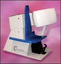 |
| Research institutions are testing the new CardiArc SPECT system from CardiArc Inc. |
CardiArc SPECT System Up for Clinical Trials
Independent research institutions will put the CardiArc SPECT system from CardiArc Inc, Lubbock, Tex, to the test. Both Cleveland Clinic, Cleveland, and Emory University, Atlanta, will evaluate the performance and design of the cardiovascular camera in clinical settings. The unit is designed specifically for cardiac imaging, particularly for coronary artery disease detection.
SPECT, or single photon emission computed tomography, noninvasively creates 3D images that show the blood flow and function of major organs, such as the heart. “Our main goal is looking at comparisons with regular patients on conventional types of SPECT imaging when looking for coronary artery disease,” said Terry Garner, vice president for sales and marketing for CardiArc Inc.
The research programs will explore how the CardiArc system stacks up to other SPECT systems. Cleveland Clinic will compare the scans of 100 patients who will be scanned by both the CardiArc SPECT system and the facility’s current clinical SPECT machine. Researchers also will compile a database of CardiArc scans that show diseased hearts as well as normal hearts both at rest and under stress. Emory University will perform similar research with a pool of 200 patients.
Both trials are expected to be completed within 6 months. “They’re really designed to show, within a clinical situation, how well does the system perform and how fast can you image?” said Garner. He adds that a faster conventional SPECT system may fall within the 12- to 15-minute range, while the CardiArc SPECT system takes approximately 3 minutes.
The CardiArc SPECT is an ultrahigh-resolution, high-contrast, upright SPECT imaging system. “We’re talking about SPECT spatial resolutions in the same realm or even better than what PET does,” noted Garner. “So, we’re talking about resolutions of down to 2 mm or 3 mm, that range.”
Once the results from the research programs are in, CardiArc will investigate broader applications for its SPECT system including the role it can play in preventing heart failure. For more information, visit www.cardiarc.com.
—Ann H. Carlson