 |
by Cat Vasko
· More Facilities Turn to IVUS for PCI extra
· Tech Zoom: Hybrid 3D Ultrasound: A Contender to CT and MR?
· FDA Clears Ashva iMagic Software
· Study Alert: “Brain Scope” Peeks Inside Skull
More Facilities Turn to IVUS for PCI extra
Coronary artery disease, which affects more than 13 million Americans, is the leading cause of death in the nation. Traditionally, angiography has been used to give physicians an understanding of the plaque buildup within arteries. Based on that information, physicians then choose a stent and plan for proper placement. However, a growing number of facilities now turn to intravascular ultrasound (IVUS), which is designed to provide a sharper image and better measurement data for more accurate stent placement.
 |
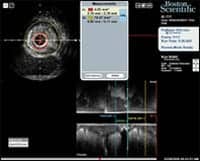
| With IVUS systems such as the iLab Ultrasound Imaging System from Boston Scientific, physicians can better analyze the type of plaque buildup, accurately size heart stents, and ensure that they are properly deployed. |
Using IVUS systems such as the iLab Ultrasound Imaging System from Boston Scientific, Natick, Mass, physicians can better analyze the type of plaque buildup, accurately size heart stents, and ensure that they are properly deployed. “We’ve seen a dramatic increase in the percentage of IVUS being used in percutaneous coronary intervention (PCI) procedures in the United States,” said Chris Japp, vice president and general manager for Boston Scientific’s imaging business. He notes that use has increased from a modest 7% to as high as 13%. “There are a number of reasons for that, but one is just the dramatic ease of use with the iLab. It’s become so much easier to use, you don’t need as much specialized training to be able to use the hardware.”
Japp estimates that approximately 600 iLab units have been integrated into facilities across the country. Although the iLab is available in the more traditional cart model, more facilities are opting for the installed version, which allows ready access to the system. “They’re integrated directly with the cath lab, so they’re always on, they’re always ready to go, and they’re very simple to use,” said Japp. “And we’ve now seen a fairly dramatic shift in sales to about 70% of the installed version of the iLab versus about 30% carts.”
Lowell Satler, MD, director of interventional radiology at Washington Hospital Center, Washington, DC, currently uses the cart version in his department, but plans are in place to equip the newest labs with the installed version. “If a cart isn’t in the room, the first question that’s asked is, ?Where is the ultrasound cart?'” said Satler. “Then you have to find it, bring it into a lab, boot it up, enter the patient data, and then connect the catheter.” He notes that ready access to an installed version will eliminate these issues.
Satler uses IVUS to assess whether pre-stent ablation or plaque modification is required to avoid inadequate expansion of the stent. “Prelesion assessment, we feel, is a very important component of IVUS, and I think that it gives you the ability to simplify the procedure,” said Satler.
The iLab’s grayscale quality, which is designed to give a clear image of plaque-deposit makeup, is the most impressive feature, according to Satler. “[iLab] has a superior image to the previous versions, larger screens, and the software interaction is more efficient and better to make measurements,” he said.
—Ann H. Carlson
Tech Zoom:
Hybrid 3D Ultrasound: A Contender to CT and MR?
Researchers at Cambridge University, in collaboration with industrial partner Dynamic Imaging, Livingstone, UK, are developing a “hybrid” 3D ultrasound system with the potential to provide data so detailed it could replace CT or MR in certain applications, according to an August 15 article in The Engineer.
3D ultrasound is currently carried out one of two ways: via a probe recording a fixed block of data, or freehand. The Cambridge device would combine the two methods, recording dense regular data like the integrated 3D probe while acquiring the larger data sets made possible by the freehand approach. The resulting system could enable physicians to clearly visualize diseased organs, and also would allow them to accurately measure the size and shape of tumors during treatment.
Because much of the information required to calculate the trajectory of the probe will be discovered by matching the blocks of 3D data, there will be no need for an inconvenient external position sensor. A miniature inertial orientation sensor will be used to guide the matching algorithms, increasing their speed and reliability, and could eventually be incorporated in the probe housing.
Now the researchers at Cambridge are focused on tracking the trajectory of the probe based on the acquired data and the output of the inertial positioning sensor. The team will also look at development and evaluation of software tools to enable the system to be used effectively in a hospital environment.
If the images are as detailed as the team hopes, they will give physicians an alternative to CT, which involves radiation exposure, and MR, which is more expensive.
—Cat Vasko
FDA Clears Ashva iMagic Software
Newly cleared by the FDA is iMagic 2.0, an ultrasound image management system from Indian PACS developer Ashva Technologies Ltd, Plymouth, Minn. The iMagic software has been approved for a variety of tasks, including ultrasound image archiving, manipulation, creation of template-based reporting, and distribution of images onto CD/DVD media. The software has been out in Asia for just over 2 years and has already racked up 500-plus installs.
“Basically, it’s reporting and distribution software,” explained Ashva USA CEO Mohan Mysore. “It runs on a standard PC or a laptop, so if somebody wants to put a laptop with an ultrasound machine and wheel it around, they can do that. It interfaces with the ultrasound machine, receives images whether they’re DICOM or non-DICOM, archives images and studies based on patient information, manipulates images and allows you to make manipulations and measurements, and prepares template-based study-specific reports in multiple formats.”
 |
| A typical iMagic screen shows the image, patient information, and image-manipulation tools together. |
Why use management software specifically for ultrasound images? “If the ultrasound machine is connected to an existing PACS, you can do everything on the PACS,” Mysore concedes. “But in large hospitals the expensive equipment is connected to the PACS. Ultrasound sometimes gets connected, sometimes does not. And many places don’t have a PACS, but would love the functionalities.”
In that sense, iMagic is much like a stepping-stone to a PACS. “We’re providing a means that’s affordable,” Mysore noted. “iMagic does everything a PACS does, at a price that is very interesting. It’s targeted for clinics that don’t want to spend so much money, but at the same time would like to get a lot of features.”
And features it has, Mysore said, detailing the list of offerings tailored especially to the needs of imaging centers. “It gives ROI information,” he said. “After using it for 6 months, it’ll tell you how many reports were normal, how many were abnormal. How many were male, how many were referred by what doctor. You can also do a cost-based study and say, I got 30% of my income from OB/GYN, 10% from general abdominal imaging, and so on. There’s a lot of such parameters fed back to the end user so he can very quickly find out where his income is coming from.”
Another interesting feature is “key images,” which enables the physician to tag an interesting image and add a footnote. iMagic also allows for the creation and storage of templates, including a special module for OB/GYN that makes it possible to make templates by trimester.
“It distributes images to a CD with a built-in viewer,” said Mysore. “A lot of people now get a CD of images instead of film, but that has not really happened yet in ultrasound. We’re hoping to make it a standard by providing a means that’s affordable.”
—C. Vasko
Study Alert:
 |
| This image taken with the ?brain scope? visualizes the canine brain in 3D. |
“Brain Scope” Peeks Inside Skull
A new 3D ultrasound scanner developed by biomedical engineers at the Duke University Pratt School of Engineering, Durham, NC, shows potential for real-time imaging of the brain. The “brain scope” is inserted into the skull through a burr hole, enabling bedside evaluation of patients when CT and MR equipment is unavailable. Findings will be reported in Ultrasound in Medicine & Biology.
“First we invented high-speed 3D ultrasound for cardiac applications from outside the body, and then in the 90s we developed intracardiac echo catheters that do 3D ultrasound. Then we switched to endoscopes, and most recently we did the brain endoscope. Wherever there’s an application for 2D ultrasound, we’ve tried to develop an analogous probe for 3D ultrasound,”said Duke biomedical engineering professor Stephen Smith, PhD.
The device is a new variation on a 3D ultrasound unit developed last year by Smith’s team for imaging the heart and other internal organs during endoscopic surgeries. To make the probe more suitable for brain imaging, the team shrank it down until it required a burr hole approximately the size of a dime for insertion.
“It works just like a 2D ultrasound probe,” explained Smith, “but instead of scanning a little slice, it scans 3D volume in the shape of a pyramid, and does that at a rate of around 30 volumes per second.”
So far the team has successfully demonstrated the “brain scope” by imaging the skull of a dog.
—C. Vasko


