|
by Cat Vasko
· Case Study: Varian Trilogy at Edward Cancer Center
· Cryoablation as Effective as External Beam Radiation for Localized Prostate Cancer in Trial
· FDG-PET Predicts Lung Cancer Patients’ Response to Chemo

- Vasudha
Lingareddy, MD

Case Study: Varian Trilogy at Edward Cancer Center
At Edward Cancer Center, Naperville, Ill, clinicians are using Trilogy technology from Varian Medical Systems, Palo Alto, Calif, to more accurately plan treatment of tumors near critical structures in the body. The Trilogy device, which comes with an On-Board Imager system that generates 3D CT images on demand, gives radiologists the capability to adapt to changes in the tumor’s size and position.
“It’s basically your traditional linear accelerator with phenomenal capabilities,” said Vasudha Lingareddy, MD, medical director of radiation oncology at Edward. “It does what every other linear accelerator does, and that’s treat our palliative cases, and delivers radiation with a wide range of energies, both electrons and photons; in addition, it has the capability to do multiple things, including stereotactic radiosurgery with brain and body, delivering pinpoint precise radiation.”
Lingareddy explained that the device’s versatility is one of its primary benefits. “You can get away with big doses of radiation because you’re localizing the tumor very accurately,” she said. “It also allows us to do image-guided radiation therapy, which is our 4D radiation therapy, along with regular fluoroscopic images. We also have a third option, and that’s our cone beam CT. So on any patient at any given time, we can obtain within seconds a CT image of the tumor or critical structures in that area and ensure that if there’s been any internal movement of the organ or shrinkage of the tumor, we can immediately identify that prior to treatment, right on the table, in addition to using the system for daily imaging.”
John Fan, PhD, senior medical physicist at Edward, noted that the software worked by analyzing the new images and comparing them to the original treatment-planning CT. “We get two kinds of information,” he explained. “One is the location difference, in terms of movements and setup discrepancies; we can get that kind of information by comparing the cone beam CT image with the original treatment-planning CT so that the software gives correction to the localization information.
“The other information we get is the tumor shrinkage in the soft tissue,” he continued. “If the tumor shrinks, we see it right away, but the software correlates the image sets together so we can see exactly how much the tumor shrinks. If it shrinks too much, it will prompt us to replan the treatment.”
Lingareddy said this kind of replanning is typically done once or twice based on the progress of the therapy. “I just replanned a patient today who had more than a 50% reduction about a third of the way through her treatment, and so in order to spare her spinal cord and her lungs, we were able to replan,” she said. “But it’s not the majority of patients who need to be replanned, and in general we just replan once. We haven’t replanned more than twice during the course of treatment.”
Which is not to say that the capability to replan isn’t a vital one, Lingareddy stressed: “We do replan, especially if we’re close to critical structures. We’ll do the cone beam CT once every other week to assess response, and then at a certain threshold, we’ll decide it’s okay to replan, there’s been enough shrinkage.”
The grocery list of procedures that can benefit from ongoing monitoring is nearly endless. “We use it for our prostate patients; we actually put gold fiducials inside the prostate and do daily images to localize the prostate at any given time,” Lingareddy said. “We also use it for lungs, not only to assess tumor response but to localize the tumor, especially if it’s near critical organ structures. We use it sometimes for the breast, because we have the capability of doing a cine image. We certainly use it for all of our brain tumors, not only for the stereotactic radiosurgery capabilities but because many brain tumors are near critical organs and structures like the optic nerves. We use it for the pancreas, for a lot of our intra-abdominal tumors.”
Anything else? You better believe it: “We’re just starting with our body stereo program,” Lingareddy said. “So that’ll be nice to be able to use for the liver and lungs.”
Cryoablation as Effective as External Beam Radiation for Localized Prostate Cancer in Trial

- The Cryocare CS from Endocare Inc was utilized in the Donnelly study to compare cryoablation with external beam radiation.

On May 22, medical device manufacturer Endocare Inc, Irvine, Calif, announced that a randomized clinical trial of 244 men with localized prostate cancer showed that cryoablation was at least as effective at treating the disease as external beam radiation. The study was the first North American clinical trial in 25 years to enroll more than 100 patients for its comparison of two definitive prostate cancer treatments.
Another interesting finding shows that 6.6% of the cryoablation patients had positive biopsy findings after 36 months?compared with 26.3% of the patients who underwent radiation therapy. Results of the trial were presented by leader Bryan J. Donnelly, MD, a urologist at the University of Calgary, at the annual meeting of the American Urological Association (AUA), Linthicum, Md.
“This study demonstrates that cryoablation is equivalent to external beam radiation when used to treat localized prostate cancer,” said Donnelly. “Furthermore, the positive biopsy rates 3 years after treatment were significantly higher for patients who underwent radiation therapy. A positive biopsy alerts a physician to recurrent or residual disease within the prostate and indicates that additional treatment may be necessary. Like radiation therapy, cryoablation is a treatment option that should be considered by all patients who are diagnosed with localized prostate cancer.”
Cryoablation was the subject of three other studies presented at the conference, and the AUA is offering a number of postgraduate courses and workshops featuring training and information on the use of cryoablation.
“The continued production of quality data by leading physicians and the presence of AUA-sponsored cryoablation courses at this year’s annual meeting mark the emergence of the cryoablation technology in the mainstream of urology,” said Endocare CEO Craig Davenport, “and indeed all areas of medicine where minimally invasive treatments for cancer are needed.”
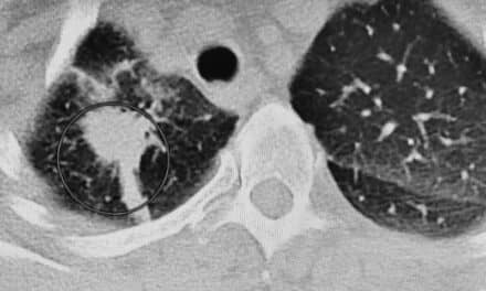
FDG-PET Predicts Lung Cancer Patients’ Response to Chemo
According to a study in the May edition of the Journal of Nuclear Medicine, PET imaging can offer clinicians an earlier indication of whether chemotherapy will benefit non-small cell lung cancer patients. The study, titled “Time Course of Early Response to Chemotherapy in Non-Small Cell Lung Cancer Patients with F-FDG PET/CT,” follows 15 patients through two courses of docetaxel and carboplatin over a 7-week period.
“With non-small cell lung cancer, since the relatively modest increase in survival must be balanced against the toxicity of the chemotherapeutic treatment, the case for monitoring therapeutic response is especially compelling,” said lead author Claude Nahmias, PhD, professor of radiology and medicine at the University of Tennessee Medical Center, Knoxville. “In spite of the persuasive findings of several studies investigating PET for monitoring response to cancer therapy, until now no published reports have clearly demonstrated that PET results were used to alter treatment.”
Over the course of the study, eight patients were classified as nonresponsive to treatment; all of them died within 35 weeks of the end of the study. Seven patients were classified as responders; of these, five survived and two died, one at 25 weeks and the other at 76 weeks. In conclusion, the researchers wrote, “Patients with non-small cell lung cancer who had a positive outcome, as exhibited by prolonged survival, were those who showed a tumor metabolic response assessed using weekly F-FDG PET studies. F-FDG PET studies performed at 1 and 3 weeks after the initiation of chemotherapy allowed prediction of the response to therapy.
“Our study demonstrates that patients who respond to chemotherapy can be identified early in the course of their treatment, and these patients will generally exhibit prolonged overall survival,” Nahmias said. “Although we studied a relatively small number of patients?and our results should be interpreted with caution?it is clear that a repeat PET study with the radiotracer FDG at the end of the first cycle of chemotherapy would allow the identification of those patients for whom the therapy was futile.
“The ability to provide an early indication of therapeutic response has the potential to improve patient care by identifying those patients who do not benefit from their current treatment,” he continued. “Patients would benefit from either having chemotherapy and its associated toxic side effects stopped or going on to a different, and hopefully more adequate, therapeutic approach.”
Finally, Nahmias noted, “Our result?that PET studies 1 and 3 weeks after initiation of cancer therapy can predict success or failure of the therapy?should be validated in a larger study in which patients are enrolled with the intention of applying it in patient management. I am forever grateful to all the patients who came back week after week to undergo our PET scans.”