|
· Philips EP Technologies Facilitate Accurate Navigation
· Tech Zoom: Beekley Introduces New Nipple Markers for Digital Mammo
· Mobile DR System Displays Images in 3 Seconds
Philips EP Technologies Facilitate Accurate Navigation
Two new navigational technologies from Philips Medical Systems, Andover, Mass, facilitate improved workflow in interventional x-ray fluoroscopy. By pairing with Stereotaxis Inc, St Louis, Philips Medical can offer magnetic catheter control; the company’s EP Navigator provides the road map, enabling practitioners to perform interventional procedures from a safe distance, sequestered from radiation inside a separate control room. Medical Imaging spoke with Frans Venker, director of development for Philips Medical’s cardiovascular business, about the two systems and how they will be integrated in the future.
 |
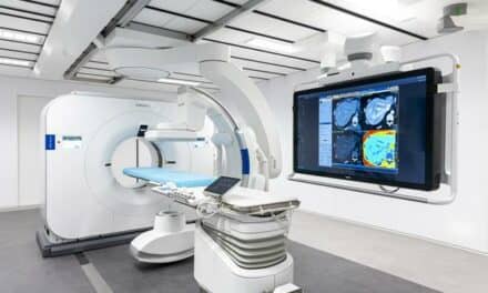
| The Allura Xper FD10 from Philips Medical has been integrated with the Niobe II from Stereotaxis. |
MI: Why did Philips Medical partner with Stereotaxis?
Venker: Stereotaxis was approached because we saw their technology for remote navigation evolving over the past few years. Research facilities have developed an interest in steering catheters without standing next to patients or within the radiation. So, the technology that came from Stereotaxis was one of the furthest-developed technologies currently available in the industry, and that’s why we made an interface between our x-ray system and the Stereotaxis system, the Niobe.
MI: How do the two technologies work together?
Venker: Actually, it’s not very simple to make them technically compatible. First, the Stereotaxis system works with magnetic fields, and magnetic fields have an effect on almost all of the components and electronics within the x-ray system. The magnet can even steer the x-ray beam, and a lot of the electrical components are affected by the magnetic field. For example, stainless steel components are attracted by the magnetic field. So, technically, a lot of adaptations had to be made in the x-ray system in order to make it magnetically compatible. On top of that, the physical size of the magnet itself gives a constraint to how the system can move; so, from an operational standpoint, software needed to be updated in order to make sure that the systems do not collide and that they know where each one is in three-dimensional space.
MI: That sounds like a long process.
Venker: Yes, it was. In short, I think it’s a process to ensure that the systems are magnetically compatible and that they don’t collide, that they work seamlessly together. It’s all workflow-related, of course, so that the customer is not hampered by any operations during the procedure.
MI: Have the initial installations gone well?
Venker: Actually, the first sites are very pleased with the system, primarily with the reliability of the system. It’s very well thought out and nicely engineered. But also from a workflow perspective, it has been designed with the customer in mind and how it will be used. Especially for atrial fibrillation procedures, it’s a wonderful addition to improve workflow to simplify the cases, which are very complex and difficult over time. So, if you talk to, for example, St Elizabeth’s Hospital [Boston], they are extremely happy with the combined installation. In fact, they indicate to us that they use it in almost 40% of the cases. That’s a lot.
 |
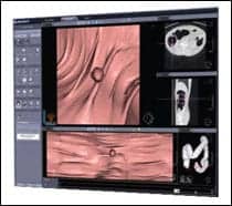
| The EP navigator is Philips Medical?s new imaging tool for complex catheter ablation procedures. |
MI: What prevented this kind of navigational technology from being possibile before?
Venker: Primarily, the technique for steering catheters within the body was always done by hand. It’s a technique that, essentially, interventional cardiologists and electrophysiologists are being taught when they learn their fields. And so the technologies to remotely steer a catheter are very limited, and Stereotaxis is by far the most advanced at the moment. They have a very complicated technology in their magnet to create a uniform field in order to steer their catheter. There are other industries and other technologies as well. Some do it robotically, with wires. But I believe those systems are still in the research phase.
MI: How does the EP Navigator differ from the Philips-Stereotaxis solution?
Venker: The Stereotaxis system is very much related to steering a catheter, so if you would like to point a catheter in a certain direction, it can facilitate that movement. The Navigator provides information about where the catheter is related to the anatomy of the heart. It doesn’t steer the catheter; it only tells you where to position it. If you incorporate a CT image of the heart into the x-ray system, the fluoroscopy is projected over the CT map. So, a clinical user sees the catheter related to the heart vessel wall. He knows where he is in the heart by steering the catheter. It’s like a GPS navigation system in a car.
MI: So, theoretically those two could be used together.
Venker: Yes. The Stereotaxis system is steering, and the Navigator is knowing where you are. The next step is to bring these technologies completely together. Based on the EP Navigator information, you know where the catheter is in space, and using the Stereotaxis system, you’re steering it wherever you would like, so that it is completely merged from a technology standpoint. For the customer, it will improve workflow even further and make atrial fibrillation cases even easier to plan. Currently, the atrial fibrillation cases show a lot of variations in the duration of treatment, so some cases could take 30 minutes and some could take 6 hours. These tools will remove that variability. For hospitals, it also will improve their planning capability.
MI: Where are you in the process of combining these technologies?
Venker: We already have this at St Elizabeth’s Hospital. They have the EP Navigator there and are researching the Stereotaxis system, and most likely, that’s where the combination will come to fruition the earliest.
MI: What are your release expectations for the combined solution?
Venker: I would not give a date yet, but it will be at least another 12 months out.
Cat Vasko is associate editor of Medical Imaging. For more information, contact .
Tech Zoom: Beekley Introduces New Nipple Markers for Digital Mammo
 |
| Designed with digital mammography in mind, Beekley?s N-SPOTS nipple markers are available in three styles. |
Last month, Beekley Corp, Bristol, Conn, introduced the latest addition to its array of nipple markers for mammography, the “Perfect for Digital” line of N-SPOTS. The N-SPOT markers feature distinct 2.3-mm nonmetallic pellets that immediately, clearly, and consistently identify the nipple’s location on the screen, without creating artifacts.
“Nipple markers continue to provide benefit on digital mammographic studies, as the radiologist can quickly and accurately determine the nipple-to-lesion distance,” Gary Whitman, MD, associate professor of radiology and associate radiologist at the University of Texas MD Anderson Cancer Center, Houston, said in a press release. “In digital mammography, placement of nipple markers eliminates the need to adjust the window and level settings.”
N-SPOTS are available in three styles: round disc, extended tab with adhesive-free center, and Soft ‘n’ Stretchy pinch-free material with adhesive-free center. For additional information, visit www.beekley.com.
Mobile DR System Displays Images in 3 Seconds
Shimadzu Medical Systems, Torrance, Calif, recently introduced its MUX-100D Mobile DaRt digital radiographic mobile x-ray system. Mobile DaRt features a 14- x 17-inch digital flat-panel detector; at just 23 mm thick and weighing 4.8 kg, the detector covers a wide examination region without inhibiting the mobility of the DaRt.
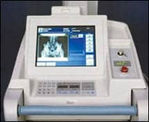 |
| The Mobile DaRt from Shimadzu offers mobile DR with an easy-to-read LCD and storage of up to 2,000 images. |
Three seconds after exposure, a reference image is displayed on the Mobile DaRt’s LCD—in other words, images can be validated immediately; image processing also can be conducted on the monitor. The device’s extralong arm features a long stroke of up to 1,200 mm, facilitating difficult imaging situations. Also, a high-frequency inverter—operating at up to 60 kHz—helps the system generate low-ripple, stable x-rays for faster and more accurate radiography.
Other features include simple network and printer output in DICOM format; storage of up to 2,000 images; intuitive maneuvering; “inch-mover” buttons on the front of the collimator for safe positioning; and clear display of x-ray timing via a bright, easy-to-read display.
For more information, visit www.shimadzumed.com.