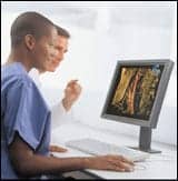
Calibrate Me
File Transfers: Easy Does It
Displays Remain Consistent Over 18-Month Watch
Product Showcase: Orthocrat Introduces TraumaCAD 3D for Surgical Planning
Product Showcase: New Visualization Software Packages Offer Speed and Efficiency
Calibrate Me
 |
| Barco?s Nio line is available in 2-, 3-, and 5-MP resolutions. |
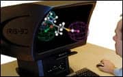 |
| A zero-cross-talk display, the IRIS 3D does not allow any of the image meant for the left eye to ?leak? into the right eye, and vice versa. |
A roundup of monitor and display releases at RSNA 2006
RSNA 2006 was the stage for showcasing a host of new monitors offering users 3D vision, side-by-side veracity, and self-calibration.
For starters, Barco, Kortrijk, Belgium, has enhanced its Nio grayscale line with a new design and a nonreflective protective front glass. Available in 2-megapixel (MP), 3-MP, and 5-MP configurations, the Nio line is bundled with ATI display controllers featuring FireGL technology?ideal for 3D imaging. All of the Nio displays have UL 60601 approval, and the 5-MP display has FDA clearance for mammography; they display 1,024 shades of gray.
Barco also showcased its MediCal QAWeb service, which the company expects to release in Q107. The service controls and monitors all displays across a facility to ensure maximum availability and compliance with DIN-6868-57, AAPM TG18, or other quality guidelines. DICOM calibration and quality-assurance tasks are performed automatically in the background without interrupting the user. If an issue occurs, the system notifies the correct person. MediCal QAWeb checks displays every 2 minutes and provides a monthly activity report for facilities; budget reports are available as well. The service comes in three service levels with varying warranties.
Next, IRIS-3D Ltd, Glasgow, Scotland, showcased its IRIS glasses-free 3D display, which presents imagery on the XRT workstation for radiation oncologists from Anatom-e, Houston. The IRIS is a zero-cross-talk stereo display that offers sharp imagery in concert with visual comfort and ease of use. With the XRT software, radiation oncologists can incorporate 3D to PET/CT, CT, MRI, ultrasound, and other modalities. Providing a standard 1600 x 1200 pixel resolution (UXGA), the IRIS can be tailored to provide dual-QUXGA-W, or 18MP. The display?s LCD panels offer 300 cd/m2 luminance and an 800:1 contrast ratio on a 20-inch window.
 |
 |
| NEC?s LCD2690WUXi is a 26-inch wide-screen offering X-Light Pro, which keeps the backlight at a constant brightness level. | The SCD 21310 from Siemens Display uses open standard video communication, allowing use of nonproprietary graphics cards. |
NEC Display Solutions of America Inc, Itasca, Ill, showcased many displays, including its new wide-screen line geared toward the operating suite and teaching. For radiology, the MD205MG-CB is a 20-inch 5-MP grayscale diagnostic display that offers a 600:1 contrast ratio; luminance is 850 cd/m2 at its maximum and 400 cd/m2 calibrated. The display features X-Light technology, which enables users to compare two display images side by side with the confidence that both monitors have the exact same brightness. Also, for reviews and referrals, NEC launched the MultiSync LCD2490WUXi and LCD2690WUXi wide-angle color displays featuring 24-inch and 26-inch views, respectively. Both offer an 800:1 contrast ratio and 400 cd/m2 luminance; both contain 1920 x 1200 native resolutions, or 2MP.
Next, Planar Systems Inc, Beaverton, Ore, enhanced its Dome EX line with increased resolution, scalability, and usability. Called the Dome DA4 architecture, it displays up to 1,024 shades of gray. Also, Planar ergonomically reconfigured the Dome E4c to help optimize reading efficiency. The EX line features the Dome E5 (a 5-MP display with 850 cd/m2 luminance and a 600:1 contrast ratio) and the Dome E4c (a 30-inch, 4-MP display with 300 cd/m2 luminance and a 600:1 contrast ratio).
The Angiography, Fluoroscopy, and X-ray (AX) division of Siemens Medical Solutions, Malvern, Pa, launched a 3D stereoscopic display geared toward angiography. The 3D display creates two or more views of an object to produce a stereoscopic impression that delivers depth information, enabling physicians to understand complex vessel structures. No special glasses are needed to use this 3D display, and viewing is not limited to one user.
 |
 |
| Siemens Medical?s 3D stereoscopic display is useable in sterile environments or in meeting rooms. | Totoku?s ME253i2 allows DICOM calibration from a remote management terminal with the same network. |
Next, Siemens Display Technologies, Alpharetta, Ga, released the SCD 21310 3MP, a color, flat-panel display with 1536 x 2048 resolution, a 400:1 contrast ratio, and 400 cd/m2 luminance. Features include an integrated stability system sensor; embedded DICOM-compliant look-up tables; and compatibility with SMfit ACT, the company?s quality-assurance software. Calibration data on the monitor is stored directly in the display, so recalibration is not necessary when workstations, graphics controllers, or other components are replaced. Siemens Display offers a constant luminance guarantee over a typical service life of 10,000 hours, or about 5 years; the backlight of the 21310 3MP has a service life of about 50,000 hours.
Finally, Totoku, Tokyo, showcased its ME253i2 and ME355i2 displays, which have an integrated luminance-stabilizing system and PM Medivisor, the company?s performance monitoring software. The ME253i2 is a 2-MP, 21.3-inch display, and the ME355i2 is a 3-MP, 20.8-inch display. Both monochrome LCDs will be released this spring.
—A. Lucas
File Transfers: Easy Does It
Two companies partner to provide another option for online imaging services
By Dana Hinesly
One paradox in today’s medical environment is that in order to succeed, small imaging centers and health care facilities must partake in available technology. However, the very nature of these organizations often means that advanced solutions are fiscally not feasible.
NearMed, Indianapolis, is hoping to change that. The company’s services, based on an application service provider business model, are aimed at reducing the costs associated with securely transferring digital images via the Internet to specialists for diagnosis and interpretation.
“NearMed offers a hosted teleradiology platform that connects rural hospitals and imaging centers with subspecialty radiologists based in major metropolitan areas to facilitate reading and interpretations,” says NearMed COO Tom Bailey. “We can either supplement existing systems or provide entire radiology coverage for these facilities.”
 |
 |
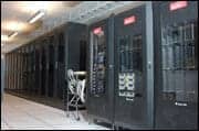
| The network operations center at n|Frame?s data center facility in Indianapolis is where NearMed?s servers are hosted and its technology infrastructure is monitored and managed. The center is staffed 24/7 to ensure constant system uptime and security. |
Such connections are made possible with a secure network infrastructure designed and managed by n|Frame, Carmel, Ind, a privately held SAS 70 Type II compliant organization. A recently announced contract between the two companies has n|Frame providing NearMed with a HIPAA-compliant method of connecting remote health care facilities with radiologists in larger metropolitan areas for the next 3 years. In other words, n|Frame is a technology company that provides the infrastructure to enable NearMed to facilitate teleradiology.
In addition to providing the ability to deliver large-format radiological images and allowing hospitals to maintain and store such records, the infrastructure enables clinicians to conduct live videoconferences over the Internet.
“We use our front-end services—which include routers, firewalls, commercial Internet connectivity, and server co-location—to provide NearMed with a network infrastructure,” explains n|Frame COO Robert Alcorn. “We also monitor NearMed’s servers and keep an eye on its capacity planning, or CP utilization, which entails monitoring about 10 different metrics to guarantee that the server continues to operate properly.”
Finding this type of comprehensive technology partner was key for NearMed. During the vendor-selection process, NearMed sought solutions with three primary requirements. In addition to wanting a commercial data center with the technological horsepower to move and store massive files from a variety of modalities, the start-up company was committed to preserving the security and confidentiality of the patient images.
“We needed to make sure the technology was well taken care of and in an environment that was managed,” Bailey says. NearMed’s preference was 24/7 monitoring of the system. “That was essential to us, because we provide service-level agreements to our clients guaranteeing its reliability and uptime.”
They found this comprehensive approach to network oversight with n|Frame, which has engineers on-site around the clock at the company’s secure, 40,000-square-foot data center. Staff of a certain caliber is a necessity in Alcorn’s view.
“We have chosen to hire actual engineers to be on staff 24 hours a day, 365 days a year. If there’s a problem, they don’t have to call someone and wait for that person to arrive; they can start the triage process immediately,” he says. “We are actively monitoring not only the system we have on our premises, but also the circuits running between us and the customer target through a virtual private network.”
Every 30 seconds, n|Frame’s monitoring systems “ping” the remote sites. If two consecutive pings remain unreturned, a technician is alerted to begin determining the extent of the problem.
Another reason the vendor stood out from the crowd for NearMed was that n|Frame did not have to start from scratch. The company accommodated the application that NearMed had developed already. Incorporating an organization’s existing technologies means that n|Frame’s clients are not required to invest any additional capital in order to take advantage of Web-based communication.
“Depending on what the facility’s requirement is, we offer choices on how they work with us,” Alcorn says, noting that his company does not manage software licenses. “We can provide a turnkey solution, which allows us to put in all the network infrastructure components, as well as servers, or they can choose to put their own hardware into our facilities and utilize our network.”
NearMed was formed and funded in the last quarter of 2005; its first client—Monroe Hospital in Bloomington, Ind—came online in September. NearMed’s co-founders—Bailey, Shyam Desiga, and Brad Bostic—predict their clients will transfer more than 60,000 large-format radiology files. They believe the product’s user-friendly aspects will help to bring in new clients.
“Users find the solution efficient and easy to use. With a fast implementation and short learning curve, productivity gains are seen very quickly,” Bailey says. “NearMed also offers training to new facilities on using the PACS in a teleradiology workflow.”
For Bailey, the bottom line goes beyond just return on investment. “[The lack of] this kind of technology has been a barrier to enabling teleradiology in many places, and we see the need that a lot of rural locations have—where they don’t have access to a world-class technology solution,” he says. “This allows them to get into a solution that is worth millions, making it possible for them to utilize technology they can’t afford otherwise.”
Dana Hinesly is a contributing writer for Medical Imaging. For more information, contact .
Displays Remain Consistent Over 18-Month Watch
During a press conference at RSNA 2006, Siemens Display Technologies, Alpharetta, Ga, highlighted the results of a study involving its displays using the Fully Automated Stability (FAS) approach to calibration. “[FAS] uses a center sensor for auto stabilization of luminance and external verification of sensor performance,” explains Ken Crocker, account manager of display technologies at Siemens Energy. “We believe that all of these elements are necessary for the successful design of a display system so that you are able to maintain consistency over time and also have consistency from display to display.”
 |
| Figure 1. The change in luminance over time for 51 3-MP displays. |
Data was collected on 51 Siemens Display monitors in the clinical setting. The displays were in use for about 18 months and had up to 8,000 hours of on time. All displays were initially calibrated at an Lmax of 400 cd/m2 and an Lmin of 0.66 cd/m2 to the DICOM Part 14 Grayscale Display Function. Luminance was measured at a number of points on the AAPM TG18 test patterns, and they were measured at routine intervals of about every 2 to 3 months.
“There was no statistically significant difference in the luminance of the 51 displays over the life of the study,” Crocker says, noting the same for brightness. “We’re very pleased to see that over the 18 months, there was very consistent display-to-display performance. So, a physician can go from one workstation to the next and expect to have a consistent image.”
Based on the results, Siemens Display determined that its monitors could be used typically about 11,000 hours before calibration would be needed. “If you assume 12 hours a day, 5 days a week, with 50 weeks in a year, that’s about 4 full years before displays would need to be calibrated,” Crocker notes.
In the end, Crocker believes that Siemens Display’s monitors are on the right track. “Some people talk about not needing to calibrate displays, but we believe that every measurement system is subject to potential for drift,” he says. “If you do not use an external photometer to at least occasionally check the performance of your display, then you run the risk of your displays drifting off into who knows where. So, it’s a design philosophy that we’ve chosen to embrace.”
Siemens Display put together a white paper based on the results. To read it in full, visit www.automation.siemens.com/monitors-med to download it as a PDF.
-A. Lucas
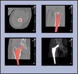
Product Showcase: Orthocrat Introduces TraumaCAD 3D for Surgical Planning
 |
| Orthocrat?s TraumaCAD 3D is now available in a Web-based, thin client model. |
At RSNA 2006, Orthocrat Inc, Petah-Tivka, Israel, showcased its TraumaCAD 3D Suite, an orthopedic surgical planning platform boasting fully functional Web-based digital templating. TraumaCAD enables DICOM-compliant importing and exporting of PACS files, including x-ray, CT, and MR images, from a local workstation or central PACS infrastructure. A variety of software tools is then available to facilitate fast, digital surgical planning.
One feature unique to TraumaCAD?s software suite is automatic calibration. Templating can be difficult when imported images are inaccurately calibrated; the TraumaCAD system both scales images imported from PACS and automatically provides calibrated on-screen implant images. By allowing different implant scenarios to be simultaneously stored and compared, the system facilitates the selection process. TraumaCAD also stores information on different vendors? implants and automatically fills out a form requesting the selected device.
Other software tools included with TraumaCAD can visualize the reduction of fractures, determine the axis of deformities, and simulate osteotomy procedures. The joint-replacement tool allows surgeons to virtually evaluate the anatomical alignment postoperation of different surgical scenarios, such as cutting, displacing, or implanting. TraumaCAD also produces anatomical measurements with an array of pediatric measurements, including growth multiplier, acetabular index, mal-alignment, spinal deformity, and more.
Now available in a Web-based, thin client model, TraumaCAD 3D automatically stores patient information, images, and procedure plans in the patient?s folder. A button is added to the PACS viewer application to launch TraumaCAD with the relevant image, and images can be transported from PACS to the planning software via drag-and-drop with PACS solutions from FUJIFILM Medical Systems USA, Stamford, Conn, (Synapse); and Dynamic Imaging, Allendale, NJ, (IntegradWeb).TraumaCAD?s template library is updated automatically, as are software upgrades. Visit www.orthocrat.com for more information.
—C. Vasko
Product Showcase: New Visualization Software Packages Offer Speed and Efficiency
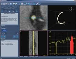 |
| Medis? QAngio CT quickly analyzes high-volume CTA data sets. |
 |
| Calgary?s ResolutionMD offers 2D and 3D visualization capabilities from any location. |
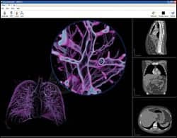
Two new applications expand the power of image processing for faster, more efficient visualization. Medis Medical Imaging Systems, Leiden, the Netherlands, offers its new QAngio CT software for detection of vessel abnormalities during CT angiography (CTA) post-processing; and Calgary Scientific Medical Group, Calgary, Canada, has announced FDA 510(k) clearance for its ResolutionMD system, which extends advanced visualization capabilities from the radiology workstation to standard computer systems.
The QAngio CT system quickly analyzes high-volume CTA data sets to offer rapid visualization independent of the CT scanner brand or type. The software features tools to automatically calculate stenosis degree, stenosis length, and an array of vessel-segment data; manual or automatic determination of stenosis and reference segments; MPR, curved MPR, and 3D views; DICOM-compatible export of analysis results in PDF; and full PACS connectivity. QAngio CT is available as an integrated or stand-alone solution, and it incorporates a floating license mechanism for efficient use of licenses by multiple users.
ResolutionMD enables standard desktop and laptop personal computers to adapt CT, MR, PET, and other medical image data from scanning systems into dynamic 2D and 3D visualizations. Offering radiologists and referring physicians the ability to perform 2D or 3D interpretations from anywhere, ResolutionMD accelerates workflow by facilitating faster diagnosis. The system works by leveraging advanced gaming graphic capabilities to manipulate and display large data sets using the computing power of both standard Microsoft Windows and MAC OS X operating systems.
“Our mission is ‘3D for every MD’—to make medical image data easy to explore and universally accessible to referring physicians as well as radiologists when and where they need it most,” Byron Osing, MD, chairman of the Calgary Scientific board, said in a press release. “Current solutions are proprietary, inflexible, and expensive, and most lack the resources to keep up with workflow requirements. We developed a cost-effective visualization tool that simplifies and accelerates diagnostic interpretation and, most importantly, moves it from the confines of the specialized turnkey workstation directly to the point of care.”
In addition to enabling interpretation on personal computers or laptops, ResolutionMD also uses its graphic capabilities to deliver real-time 3D rendering and contextual close-up capabilities for the exploration of obstructed anatomy without cumbersome volume segmentation. An interactive lens tool and an edge-enhancement tool improve visualization of low-contrast internal structures. To learn more about ResolutionMD, visit www.calscimed.com; for more on QAngio, visit www.medis.nl.
?C. Vasko