Running the Numbers
DIVO: Quick Fix for Digitizing Echocardiograms
Associations Unite to Improve Quality of Thyroid and Parathyroid Ultrasound
Taking Note of Transducer Technology
Study: Portable Ultrasound Superior to FRS for Evaluating Heart-Disease Risk in Women
Running the Numbers
1 is how two different surveys regarding ultrasound ranked both GE Healthcare, Waukesha, Wis, and Philips Medical Systems, Andover, Mass.
GE Healthcare was ranked #1 by IMV Ltd?s 2006 ServiceTrak survey in 13 key performance categories for Radiology/OB Ultrasound Service, including effectiveness in resolving problems, remote-service diagnostic efficiency, and overall service performance relative to change. The ServiceTrak survey also ranked Philips Medical #1 in overall service performance for Patient Monitoring Systems and Ultrasound All Systems, comprising Radiology/OB and Cardiology instruments. Finally, MD Buyline?s third-quarter 2006 survey ranked GE Healthcare #1 in system performance, installation and implementation services, and service response time for Radiology Ultrasound.
ServiceTrak by IMV Ltd, Greenbelt, Md, is a survey of respondents drawn from a randomized database of US hospitals and imaging centers; for more information, visit www.imvlimited.com. MD Buyline, Dallas, maintains an online database of medical technology and informatics products, price, and performance; its results are based on, among other things, user-satisfaction surveys and interviews. For more information, visit www.mdbuyline.com.
DIVO: Quick Fix for Digitizing Echocardiograms
By Dana Hinesly
In an era when most people no longer use VHS tapes, it seems somewhat perplexing that ultrasound technology still relies on them. “The traditional protocol of sending a videotape via a courier was unbelievable to me,” says Don Wauchope, creator of digital images from video output (DIVO), which is a simple medical device that connects to an ultrasound machine and digitizes echocardiograms. “I would ask these doctors why they were continuing to do that when there are digital methods of sending ultrasound that have been around for years.”
The answer is straightforward. Ultrasound captures moving pictures and, when constrained to 5-second loops, clinicians don’t have the information they need to make a confident diagnosis. As it turns out, the solution is just as direct.
Once captured, technologists and clinicians have an electronic file to do with what they will. Most DIVO users transmit the footage to a physician via a virtual private network, using file transfer protocol software. The system defaults to produce uncompressed images, but users can make adjustments as they deem appropriate.
Currently in use at more than 50 facilities around the world, including hospitals in Guam and Holland, the FDA-approved device seems to be having the biggest impact in the lives of pediatric cardiologists, whose patients often defy convention.
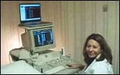
 |
| Sonographer Linda Cabrera uses the DIVO system mounted on a Vivid ultrasound system from GE Healthcare. |
“Adults always have the same heart problems, but babies are always different,” Wauchope explains. “Although there are some standard conditions, more have unique problems. The liability is so high, doctors can’t make a diagnosis from just a few frames.”
As a result, pediatric echocardiography specialists require long, extensive studies that sweep very slowly through the heart. Audio plays a vital role, because often defects can be heard but not seen. “These are all things that are really not that necessary with adults, but are crucial with children,” Wauchope says. “DIVO can capture those extended-length videos and audio, [including Doppler and technologists comments,] as well as still-motion images.”
Capturing the image stream does not require any special adapters. The DIVO device connects to the same S-video and audio outputs used to record from the ultrasound machine onto videotape.
Installing the solution does not limit the technologists’ options. The ultrasonographer can record directly onto the DIVO, onto the VCR, or into the ultrasound machine’s hard drive for future transfer to the DIVO. Viewing does not require proprietary viewing software. All image files from DIVO can be viewed on any standard PC running Windows.
It also is important to note that the PC does not need to be a desktop. Wauchope recounts an experience that one of his clients had recently. The pediatric cardiologist received an urgent page when she was more than 20 miles from the emergency department that was treating the infant. Using her laptop computer, which had a mobile broadband card installed, she was able to view the echo results before arriving at the hospital—an act she believes saved the child’s life.
Adds Robert D. Loitz, MD, the physician’s colleague at Pacific Pediatric Cardiology Medical Group, Los Angeles, “[DIVO] eliminates the delay we would invariably have covering acute care nurseries ? in being able to immediately diagnose babies with often life-threatening heart disease,” he says. “The echo is coming to us at the speed of light, [and] that can be the difference between living and dying.”
Not only is the DIVO faster, but the resulting image trumps VHS quality. “It’s vastly superior to videotape, which is the traditional medium,” Wauchope says. “It’s the same as what they see on the screen.”
Another difference between DIVO and other advanced technologies is that the former won’t break the bank. “We sold one to the University of California, San Diego, and the doctor had a difficult time buying it because it didn’t cost enough,” Wauchope says. “That’s actually what they said. It got all the way up through purchasing and got bumped back because they had just spent a quarter of a million dollars on an OEM system that couldn’t do what this does.”
Wauchope collaborates with Pyramid Medical Inc, Los Alamitos, Calif, to manufacture and distribute the device and is actively seeking distribution channels for the DIVO product. Although pediatric cardiology has ignited Wauchope’s passion, DIVO can be used for any specialty.
Dana Hinesly is a contributing writer for Medical Imaging. For more information, contact .
Associations Unite to Improve Quality of Thyroid and Parathyroid Ultrasound
The dissemination of diagnostic ultrasound continues as the American Institute of Ultrasound in Medicine (AIUM), Laurel, Md, and the American Association of Clinical Endocrinologists (AACE), Jacksonville, Fla, have announced their joint commitment to enhance training for thyroid and parathyroid ultrasound procedures. Per the agreement between the two associations, those endocrinologists recognized for thyroid and parathyroid ultrasound by the AACE also will be recognized as having demonstrated satisfactory training by the AIUM.
“[The AACE] wanted to work with us on developing training guidelines that would be acceptable to us if a facility were to apply to be accredited by the AIUM,” says AIUM President Lennard Greenbaum, MD. “[The AACE] had a training program, but it had not done any physician certification in the past other than a certificate indicating that the physician had attended the course.”
The AACE has been conducting its course in using ultrasound to evaluate thyroid and parathyroid disorders since 1998; the curriculum includes both diagnostic ultrasonography and ultrasound-guided fine needle aspirations (UGFNA). Now, the AIUM acknowledges the revised course as worthy of accreditation. “Achieving recognition by AIUM for meeting high educational standards in ultrasound is a tremendous accomplishment,” AACE President Steven M. Petak, MD, said in a press release.
AACE’s enhanced requirements for thyroid and parathyroid ultrasonography certification and recertification consist of 15 credit hours, spread over 2 days, including both didactic and laboratory sessions. Once issued, the certification is conditional, requiring “follow-up verification of continuing activity and interpretation of images performed from an ultrasound study by an ultrasonographer (technician or physician) and the performance of UGFNA procedures over the ensuing 12 months,” according to a statement published by the AACE.
Greenbaum says, “It was a very cooperative effort. Ultrasound is changing, and with the miniaturization that’s occurring and the advances in technology, many specialties have worked ultrasound into clinical practice. We hope that the partnership that we have worked out with the endocrinologists will be a template for dealing with other specialties.”
?C. Vasko
Taking Note of Transducer Technology
RSNA provides a bevy of new transducer introductions
 |
| The high-resolution 4D transvaginal probe from GE Healthcare helps clinicians see and detect fetal abnormalities earlier. |
Transducer technology just keeps improving, as evidenced by the new releases at the 2006 RSNA Annual Meeting. More coverage areas, better resolution, and the ability to pick up subtle details are the name of the game.
 |
| For the Acuson Sequoia, Siemens Medical released two new transducers. |
 |
| SonoSite?s C11e/8-5 transducer (above left) is ideal for anesthesia and pediatrics; the P17/5-1 transducer (above right) focuses on cardiac, transcranial, and abdominal imaging. |
 |
| ZONARE?s L12-5sp transducer captured the peroneals (above), and the P4-1c captured the Circle of Willis (below). Click on either image for a larger view. |
 |
First, GE Healthcare, Waukesha, Wis, released a 4D transvaginal probe for the Voluson E8 system. According to the company, the transducer can help clinicians detect fetal abnormalities earlier than before, as well as improve diagnostic confidence in complex GYN exams. The 4D probe features a wide field of view and volume-scanning angle, both of which gather volumetric information on subtle anatomic structures.
Next, Siemens Medical Solutions, Malvern, Pa, released two new transducers for the Acuson Sequoia platform. The 17L5 HD is a high-density, ergonomic transducer that features patent-pending SureGrip for better handling. With 512 individual elements, the 17L5 provides high contrast resolution, spatial compounding, and steering angles, all of which enable great detail in breast, small parts, complex masses, and subtle pathologies. Also, Siemens Medical released the 9L4 Multi-D transducer, which is geared toward vascular imaging, including cerebrovascular, peripheral arterial, and deep venous imaging. It provides simultaneous B-Mode, color Doppler, and pulsed-wave Doppler. The Multi-D Array technology provides improved contrast and spatial resolution through uniform elevation focusing; the transducer’s Virtual Format imaging provides a wide field of view to completely display anatomy.
Third, SonoSite Inc, Bothell, Wash, released two new transducers at RSNA—along with a major upgrade to its MicroMaxx system. The C11e/8-5 is a micro-convex array that enhances visualization in regional anesthesia and line placement as well as the pediatric abdomen. The P17/5-1 phased array transducer has been enhanced for 2D imaging with clutter reduction, improved harmonics, and—keeping an eye on the growing obese population—increased imaging penetration to 35 cm. The P17/5-1 is geared toward cardiac, transcranial, abdominal, and OB/GYN imaging.
Finally, ZONARE Medical Systems Inc, Mountain View, Calif, also released two transducers. The P4-1c is designed for transcranial and abdominal vascular imaging. “For those, you need a slightly different format and a smaller footprint to get deep vessels in the abdomen, and indeed, this transducer does very well with it,” explains Lars W. Shaw, vice president of marketing at ZONARE, noting that it offers 30 cm of penetration. Measuring 14 x 21 mm, this transducer has a combination of nine multiple frequencies within 2D and M-Mode, tissue harmonics, color/power Doppler, and pulsed-wave Doppler. A work-in-progress capability of the P4-1c is cardiac imaging, which the company expects to be available at RSNA 2007. ZONARE also released the L12-5sp Linear Array—”our hockey-stick intraoperative transducer that does high-frequency imaging, from 5 to 12 MHz,” Shaw explains. Offering up to 6 cm of penetration and with eight multiple frequencies, the L12-5sp is geared toward intraoperative vessel scanning, guiding needles/catheters, musculoskeletal, superficial vein mapping, vein ablation, and soft-tissue biopsy.
?A. Lucas
Study: Portable Ultrasound Superior to FRS for Evaluating Heart-Disease Risk in Women
Risk for heart attack is generally stratified using the Framingham Risk Score (FRS), a complex equation incorporating such factors as gender, age, previous or current smoking, presence of diabetes, blood pressure, and cholesterol score. But at the World Congress of Cardiology annual meeting held September 3?6 in Barcelona, Spain, John Postley, MD, of the New York Physicians Group presented a study showing that FRS stratification frequently fails to identify at-risk women—and that ultrasound is a superior identifier of risk.
“Unfortunately, Framingham has been rather poor in identifying individual people,” Postley explains. “We have people at low risk who suddenly keel over with a heart attack. And then, we have people at high risk who seem to do all the wrong things, and they do fine. Everybody had been talking about the fact that doctors had underdiagnosed women. And the question was, can you prove it?”
 |
 |
| SonoSite?s MicroMaxx system displays thickening of the internal carotid wall, an early indicator of cardiovascular disease before plaque develops. | This image of the carotid artery bifurcation from the MicroMaxx system shows the presence of plaque on both the near and far walls of the vessel. |
In the study, 120 patients from Postley’s practice—70 men and 50 women—were examined with a Titan portable ultrasound system from SonoSite Inc, Bothell, Wash, for the presence of plaque in the carotid and femoral arteries. Measurement of carotid intimal medial thickness (CIMT), a precursor to heart disease, also was conducted. “Although Framingham Risk Score is barely better than 50% at identifying the people who subsequently will have disease, if you can find people who have disease, you can find modifiable risk factors in more than 50% of them, which will change their secondary risk,” Postley explains.
He found no correlation between the FRS and CIMT in the 50 women studied. According to the FRS, 72% of the women were at low risk for heart attack; ultrasound revealed arterial plaque, another proven precursor of heart attacks and stroke, in 50% of the group. “There was a moderately significant correlation between presence [of plaque and/or CIMT] and Framingham Risk Score in men, and absolutely no correlation in women,” he says. Postley also notes that a larger study of 2,500 women, involving cardiologists from Johns Hopkins Medicine, Baltimore, and the University of California, Los Angeles, found the same lack of correlation between FRS and precursors to heart disease—in this case, coronary calcification. “The studies were fairly conclusive,” Postley says.
With a resolution limit of one tenth of a millimeter, ultrasound is capable of identifying CIMT before CT or MR angiography; it also is useful at evaluating the progress of treatment, according to the study authors. “Our hypothesis is that although the plaque is the actual instigator of disease, when you institute treatment, you don’t see a big change in the plaque,” Postley explains. “It takes a couple of years to see a change. But if you institute a successful treatment, you’re going to find a change in the CIMT in a matter of 6 months. Look at the CIMT, and you’ll be able to see a change in that thickness, which will give you confidence that you’re on the right track.”
And with 250,000 Americans dying from first heart attacks every year, that kind of confidence could prove invaluable. “There are 44,000 women dying of breast cancer every year,” Postley says. “There are 600,000 first heart attacks each year. Every woman goes and gets a mammogram, but no one does anything about their arteries.”
?C. Vasko


