IVUS provides more information in a shorter time
Expert Tips: Self Help for Sonographers to Avoid Work-Related Injuries
Ultrasound Study Benefits US Olympic Athletes
Running the Numbers
Georgia Tech Researchers Develop Portable Vein Finder
IVUS provides more information in a shorter time; Georgia Tech develops portable vein finder; and tips for sonographers to avoid work-related injuries.
by Dana Hinesly
Very high-intensity statin therapy resulted in significant regression of atherosclerosis, according to a recent study. Beyond the therapeutic ramifications of this study—titled “Effect of Very High-Intensity Statin Therapy on Regression of Coronary Atherosclerosis: The ASTEROID Trial”—perhaps its bigger contribution is the further validation of intravascular ultrasound (IVUS) to monitor disease progression.
“Intravascular ultrasound is a very sensitive methodology to look at changes in the coronary arteries,” said Christie M. Ballantyne, MD, director of the Center for Cardiovascular Disease Prevention at the Baylor College of Medicine and the Methodist DeBakey Heart Center (Houston) and one of the study authors. Ballantyne noted that although the coronary angiograms historically used are helpful in terms of looking at the lumen, they are limited in the amount and type of information they can provide. “Through IVUS, you can still look at the lumen so that you can still quantitate that,” he said, “but you’re also getting much more about what’s happening in the vessel wall.”
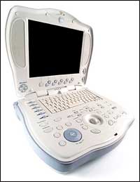
An IVUS procedure involves threading an ultrasound-tipped catheter through the coronary arteries to obtain detailed images of the atheroma within the wall, in addition to the lumen of the arteries. It allows clinicians to visualize the atheroma area and look at the percent atheroma volume, among other parameters, such as acquiring measurements of the arterial wall.
Even with its additional benefits, IVUS procedures are cost-prohibitive for widespread use in therapeutic and diagnostic capacities. However, the impressive results achieved with the technology have pushed it to the head of the pack in coronary research circles.
“This study is showing the potential of IVUS to detect the regression in human atherosclerosis in a reasonable sample size over a relatively short period of time,” Ballantyne said. “And I think IVUS has moved up as the state-of-the-art technology for measurement of the effects of new drugs on coronary atherosclerosis.”
Due to its ability to collect a more comprehensive measurement of atherosclerosis and its increased sensitivity to picking up changes in the arterial wall of the coronaries and lumen, researchers are employing IVUS to reduce the period of time for clinical trials. The amount of time saved varies by individual assessments, but shorter study times close the distance between new pharmaceutical treatments and data about their efficacy and safety. Benefits of this abbreviated cycle go far beyond bringing medications to market more quickly.
“This has major implications for getting the answers about how well a drug is working, in obtaining information for regulatory authorities, and perhaps in changing the labeling of agents with indications,” Ballantyne said.
The potential impact on patient care has generated a very enthusiastic interest in identifying other, more sensitive ways to detect changes in the coronary arteries even more rapidly. To this end, many researchers are focusing on noninvasive procedures—such as CT angiography and MRI—as an alternative. But to date, such methods have not been able to give the precision of the measurement that can be realized with IVUS.
Another area of enthusiastic investigation is the attempt to obtain even more information from IVUS than it provides today. “A number of catheters have been developed, and other research is in progress, hoping that one could even get more information by further refinement of this technology,” Ballantyne said. “A number of investigators are trying to see if it’s possible to gather more data in regard to information about what’s happening in the vessel wall, trying to see if they can identify changes from stable to unstable plaque—these are the types of investigations that are ongoing.”
The study was published in the April 5, 2006, issue of the Journal of the American Medical Association. For more information, visit JAMA (sign in page – restricted access for nonsubscribers).
Expert Tips
Self Help for Sonographers to Avoid Work-Related Injuries
by Renee Diiulio
According to surveys, the majority of sonographers are scanning in pain. The simple solution is to stop it; but room design, older products, workloads, career goals, and the need to eat prevent many from taking this advice. Product design and department policies can help, but sonographers also can help themselves. Medical Imaging gathered some tips from experts on how to do this:
- Take time at the beginning of each workday to stretch.
- Prepare the work setting to accommodate patient needs and ergonomic solutions.
- Participate in purchasing decisions; request ergonomic equipment.
- Learn to use the ergonomic equipment available.
- Stay aware of your body position, and practice good mechanics. Minimize sustained bending, twisting, or forceful movements.
- Make sure the monitor is at eye level and about an arm’s length away.
- Keep your arm as close to your side as possible and abducted no more than 30%.
- Get as close to the patient as possible.
- Lift patients with the help of others, using your legs—not your back—to lift.
- Allow the patient to assist you.
- Learn to work with both of your hands. This might require some room reconfiguration, but it can reduce the chance of injury by half.
- Take microbreaks during scanning to relieve fatigued or strained areas.
- Vary work duties throughout the day.
- Participate in an exercise program that includes targeted muscle work to protect known stress points.
- Follow general healthy practices: Eat right, get plenty of sleep, and exercise.
Looking for more information on sonography-related injuries? Don’t miss “Pain in the Neck?“.
Ultrasound Study Benefits US Olympic Athletes
 |
| Using the Logiq Book XP portable ultrasound system from GE Healthcare, study researchers are monitoring shoulder- and knee- injury assessment of US Olympic Women?s Ice Hockey Team members. |
A recent initiative by GE Healthcare (Waukesha, Wis) could transform athlete medical care and uncover health care benefits for the general public. The clinical research study—which involves the US Olympic Women’s Ice Hockey Team and focuses on musculoskeletal (shoulder and knee) injury assessment—is part of GE Healthcare’s sponsorship of the Olympic Games through 2012.
Along with project leaders Marnix T. van Holsbeeck, MD, and Scott A. Dulchavsky, MD, PhD, of Henry Ford Health System (Detroit), study researchers will investigate, over the course of a year, whether taking healthy baseline scans of all 24 members of the US Women’s Ice Hockey Team helps in determining the extent of future sports injuries with greater speed and accuracy. Researchers are using GE Healthcare’s portable Logiq Book XP ultrasound system.
“We discovered that the trainers could perform difficult musculoskeletal ultrasound examinations, with high accuracy, after just a few hours’ training,” Dulchavsky said. “We also identified a number of injuries in the highly competitive athlete population, which were not known prior to the study, but would impact treatment decisions if the athletes were injured. We also developed video-streaming solutions, which allowed real-time, remote assessment of athletes by physicians thousands of miles away.”
According to Dulchavsky, it is a challenge to evaluate injuries in the field, which makes this study useful. “Having a healthy baseline scan of common injury sites, such as the shoulder and knee, as well as portable ultrasound technology on-site, could be a step that fundamentally improves our ability to care for athletes’ injuries. Our study with the female ice hockey players could ultimately be applied to any sport, benefiting athletes from any sport or skill level,” he said.
The clinical study began in September 2005, and initial results are expected in late 2006. GE Healthcare expects to conduct similar athlete research programs in other countries later this year.
The study authors will conduct additional training/familiarization sessions with athletic trainers in anticipation of the upcoming 2008 Summer Olympics in China. They have used the techniques to diagnose injured athletes during training and hope to provide additional medical capabilities during the next Olympic Games.
Running the Numbers
40% of the 2,975 hospital-based general ultrasound or radiology departments surveyed by the IMV Medical Information Division (Des Plaines, Ill), reported that they will improve their current ultrasound capability by adding new units, replacing old ones, or updating current units.
Georgia Tech Researchers Develop Portable Vein Finder
A team of researchers at the Georgia Institute of Technology (Atlanta) is developing an inexpensive, handheld device that employs Doppler ultrasound technology to find veins quickly. The researchers hope to curb the difficulties that are encountered when dealing with trauma patients under trying circumstances.
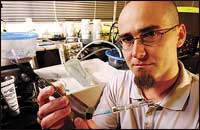 |
| A team of Georgia Tech researchers, including research engineer Francois Guillot in the School of Mechanical Engineering, is developing an inexpensive, handheld device that uses Doppler ultrasound technology to find veins quickly. |
“Depth and angle are the critical issues for vessel detection,” said project leader Michael Gray, a research engineer at the Electro-Optical Systems Laboratory within the Georgia Tech Research Institute. “Even if you locate a vein at the skin’s surface, it’s still easy to miss when you try to insert a needle into the tissue below.”
The patent-pending vein finder comprises two parts: A reusable unit houses the electronics and signal-processing components, and a disposable coupler box holds a reflector and needle guide. The needle guide is positioned parallel to the sound beam being transmitted by a transducer in the device’s reusable section.
“The device transmits sound into the skin and looks for sound scattering from flowing vascular blood, indicated by a Doppler shift,” Gray explained. “The device would indicate the presence of a blood vessel, and whether it was a vein or artery, with a simple light display, similar to that which is used in wall-stud finders, for example. The user would move the device on an arm or other area of interest until the desired vessel is located. At this point, the user can insert a needle or catheter into the vessel by sliding it along a guide that is part of the device. The device was designed so that the needle is guided along the same path as the sound beam used to detect the vessel, therefore ensuring that the needle/catheter goes to the desired location.”
The vein finder has been proven effective in initial tests on phantom tissue, a model that simulates human tissue and blood vessels. Researchers have now begun adapting the device for human tissue.
Still, developing the user-friendly vein finder has been deceptively complex. “One reason it’s so challenging is that we’re using very simple components to keep costs down,” said Francois Guillot, a research engineer in the School of Mechanical Engineering.
As for its availability, Gray said, “The proof-of-concept prototype needs further refinement. We don’t have a firm schedule at this time, but we hope to be done with the development by the end of the year.” He also noted that in addition to the device’s utility in trauma, it is expected to be applied in testing and other applications for clinical use.