A Closer Look at the NFL Combine
Collaboration: Biophan and Siemens Medical Declare Collaboration Intent on Advanced Interventional MRI
Running the Numbers
Neurognostics and Medical Numerics Join Forces for fMRI
MRI Suite Safety Found in Four Zones
Siemens, IMRIS Offer Ceiling-Mounted MRI
A Closer Look at the NFL Combine
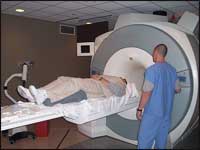
by Dana Hinesly
 |
| Mike Bortone, RT, (standing) demonstrates the 3T MRI scanner with the help of “patient” Jim Braswell, RT. |
For National Football League (NFL) hopefuls dreaming of a career under the stadium lights, the biggest hurdle to success might not be their on-field stats. Every year, the NFL invites a select group of college players for intense medical, physical, and psychological evaluations known as the NFL Combine.
The 2006 Combine marks the 20th year it has been hosted by Methodist Hospital (Indianapolis), a member of Clarian Health Partners.
“It used to be that if teams were interested in a college player, they would test him individually,” said John Dickey, radiology manager at Methodist Hospital. Dickey oversees the coordination of all diagnostic testing at the hospital and has been involved with the Combine since its beginning at Methodist Hospital. “It reached a point where a player would travel to five or six different cities, having the same tests done over and over, until the NFL finally decided to have players tested at one place and provide all teams with the same information.”
Prior to settling on Indianapolis, the Combine would travel from city to city. In 1987, the Indianapolis Colts hosted the event with such efficiency that NFL administrators decided the Combine should stay put. Since then, the week after the NFL Pro Bowl finds Methodist Hospital’s staff bracing itself for 4 long days processing athletes from across the country.
Although it started small—the first year’s Combine produced only three MRIs—the event’s growth has been substantial. This year, 330 athletes were examined, and 348 MRIs were performed.
Imaging studies account for only part of a rigorous physical to which each athlete is subjected and are performed only on individuals who have suffered an injury at some point in their football careers.
“Any time a player has any sort of musculoskeletal injury or neurological injury, we’re going to check that out very, very closely,” said Dickey, who notes that the nature of the sport translates to some sort of imaging exam being performed on almost every contender. “We very seldom do an MRI on the kickers, because they don’t have as much contact, but that’s about the extent of it, because everybody else gets pretty beat up.”
Performing MRIs on these highly conditioned men presents some unique complications. “The biggest challenge we have is that a lot of the players are just huge human beings, so we find it very difficult to get them into the scanner, which compromises our ability to perform these diagnostic tests,” Dickey explained. Many of the players also struggle with claustrophobia, so some of them are examined using one of the hospital’s open magnets.
Numeric values are assigned to the results from each exam, resulting in a cumulative score that enables the NFL team physicians and trainers to rate players, which helps determine the NFL’s professional interest in them. Because results can change the course of each athlete’s future, it is not uncommon for the MRIs, CTs, and x-rays to reveal a host of undisclosed injuries, such as knee, shoulder, neck, and back ailments.
“We also find things nobody had any idea about, something asymptomatic that could have caused problems later,” Dickey said. “Just about every year, we discover things like lung and bone tumors and renal tumors—things the athlete didn’t realize he had, but that will definitely change his draft status.”
Each year, Methodist Hospital employs 14 different 1.5T MRI systems—manufactured by GE Healthcare (Waukesha, Wis) and Siemens Medical Solutions (Malvern, Pa). But this year’s Combine helped break in the health care facility’s new Magnetom Trio from Siemens Medical, a 3T magnet that came online the week before players started arriving. The hospital’s scanner is the only 3T magnet in the state solely devoted to clinical use.
“We were hearing from our clinicians that they wanted us to upgrade to a 3T magnet, which definitely has some advantages as far as image quality,” Dickey said. For example, the Trio produces higher-resolution scans for neurophysiology studies in less time than its lower-field counterparts. “We added the 3T to provide the highest-quality imaging available to our patients.”
Collaboration
Biophan and Siemens Medical Declare Collaboration Intent on Advanced Interventional MRI
Biophan Technologies Inc (West Henrietta, NY) has announced a declaration of intent to enter into negotiations for a strategic research collaboration between Siemens Medical Solutions? MRI Division (Erlangen, Germany) and Biophan Europe GmbH. The goal is to provide new solutions in interventional MRI and MRI-assisted minimally invasive surgeries.
The collaboration is the result of discussions between senior officers at the two companies for the further development and commercialization of potentially complementary MRI technologies. Biophan is attempting to enable effective MRI visualization of implanted medical devices, including heart valves, coronary and peripheral stents, and vena cava filters. Also, Siemens Medical has developed a new MRI-scanning technology with aperture configurations that could be highly complementary to Biophan?s research and development program in this area, according to Biophan Europe CEO Michael H. Friebe, PhD.
Many implantable and surgical devices containing metallic elements are contraindicated for use with MRI due to their potentially undesirable interactions with the powerful electromagnetic fields generated by MRI systems. To extend MRI to patients with implantable devices, who currently are denied access to MRI visualization, Biophan has developed a range of proprietary technologies to enable MR imaging to visualize the interiors of stents and other implants, and to enable the MRI-guided implantation of devices.
Running the Numbers
347 current US installations of 3T MRI scanners.* The numbers include 167 installations of the Signa HD and HDx lines from GE Healthcare (Waukesha, Wis), 60 installations of the Achieva line from Philips Medical Systems (Andover, Mass), and 120 installations of the Magnetom Trio line from Siemens Medical Solutions (Malvern, Pa).
*Numbers are based on a May 11 vendor poll.
MRI Suite Safety Found in Four Zones
by Nancy Salisbury
Even with space at a premium for most hospitals and imaging facilities, finding the appropriate square footage for a successful MRI service begins with a facility layout designed around optimum patient care and safety. Today, it’s as easy as 1, 2, 3 … and 4, thanks to the MRI suite four-zone safety-guiding principles.
Established by the “White Paper on MR Safety and MR Safe Practice Guidelines” from the American College of Radiology (ACR of Reston, Va), this set of policies provides safeguards and fail-safe mechanisms to help protect patients, attending health care professionals, and anyone else who occasionally enters the magnetic field of MR scanners (such as housekeeping or security personnel).
“The four-zone guidelines are a universal set of simple fixes that can be implemented relatively easily and inexpensively to help avoid adverse affects of accidental exposure to magnetic fields by non-MR personnel,” said Emanuel Kanal, MD, FACR, committee chair of the ACR Blue Ribbon Panel on MR Safety.
Conceptually divided into the four zones, the MR suite should have progressive restrictions:
- Zone 1 is freely accessible to the general public and is just about everywhere that the site’s magnetic field is not, leaving just three zones for imaging facilities to manage.
- The reception and waiting areas generally compose Zone 2; those who enter are under the supervision of trained MR staff who thoroughly screen patients for MR contraindications.
- Zone 3 contains the control room and computers, and it should be physically restricted from general public access. Its stringent admittance should be controlled by and entirely under the supervision of MR personnel, such as the MR technologist or patient coordinator, with no exceptions to this guideline.
- The most secure and closely guarded area is Zone 4, which is synonymous with the MR scanner magnet room. It should be clearly marked as being potentially hazardous due to the presence of very strong magnetic fields.
MR personnel should have direct visual observation of the scan room from the control room, as well as all pathways between Zones 3 and 4. This safety precaution is the last barrier for MR personnel to deny access of unscreened individuals and ferromagnetic objects or equipment into the scan room.
Whether executed at the inception of an MR facility or as structural adjustments to an existing MR suite (such as strategically added doors and locks), the four zones work in unison with each other and can actually give the added bonus of improved patient throughput. For ongoing MR-facility safety, internal checks using the ACR guidelines should be conducted annually or whenever an imaging center introduces any significant changes to the MR service or environment.
Read the ACR’s report in full.
Siemens, IMRIS Offer Ceiling-Mounted MRI
Siemens Medical Solutions (Malvern, Pa) and IMRIS Inc (Winnipeg, MB, Canada) have signed a global original equipment manufacturer (OEM) agreement to incorporate IMRIS’s iMotion—the company’s ceiling-mounted surgical imaging systems that are used in neurosurgical, spinal, and soft-tissue procedures—into Magnetom Espree and Magnetom Symphony MRI Systems.
 |
| The ceiling-mounted Magnetom Espree with IMRIS iMotion is installed at three locations to date: Foothills Hospital in Calgary, Alberta; Children’s Hospital in Boston; and Wilkes-Barre General Hospital in Wilkes-Barre, Pa (shown here). |
“The magnet is suspended from the ceiling on an MRI-safe crane. The magnet can then be moved from room to room,” explained Ed Gunning, marketing manager of cardiac and interventional MRI in the MRI division of Siemens Medical. This ability allows the advanced technology to be present in multiple locations. Additionally, the combination of Siemens Medical’s diagnostic technology and IMRIS’s surgical innovations help surgeons remove diseased tissue more efficiently, reduce damage to healthy tissue, and decrease visits to the operating room (OR) for subsequent surgeries.
Added IMRIS President David Graves, “The challenge of having an MRI system solely in the OR is that it can be costly and could take medical facilities years to recoup their investment. By offering a system that can be used in the OR as well as the diagnostic-imaging department, a facility can realize a significantly improved return on investment. This need is filled with iMotion by providing advanced MRI technology in either department at any time, in minutes.”
IMRIS originally approached Siemens Medical for this partnership due to Siemens Medical’s technology and system design, according to Gunning. The Espree became the system of choice because of the scanner’s large opening (70 cm). The testing took place mostly in IMRIS’s factory in Canada with assistance from Siemens Medical’s staff.
—M. Said


