 |
Heart disease has long been the number one killer in America—but recent statistics from the Centers for Disease Control and Prevention (CDC) give new hope. In January, the CDC reported that deaths from coronary heart disease declined 25.8% between 1999 and 2005. Based on these numbers, the American Heart Association (AHA) projects that 240,000 lives will be saved in 2008 alone. (To learn more, read: AHA News – January 22, 2008)
While the AHA reports that Americans must take immediate steps to reduce risk factors for heart disease if these numbers are to hold, the association credits ongoing advances in medicine, technology, and evidence-based research for the improved survival statistics. As a vital part of this momentum, 3D cardiac imaging will continue to play a prominent role in helping physicians detect and treat heart disease.
Number One 3D Tool
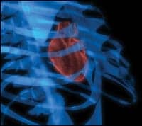
Images of the heart were once difficult to capture, because the organ moves in more than two planes. But today, radiologists have an arsenal of imaging modalities at their disposal to get a clear understanding of exactly where heart disease is located and how far it has progressed.
 |
MR and ultrasound, long staples of the trade, provide excellent insight into heart size, function, and valve abnormalities—but only CT has the spatial resolution to allow radiologists to see into the coronary arteries themselves. In the past several years, 3D CT technology has evolved rapidly, and today’s machines offer radiologists better clarity and faster results than ever before.
“The other [modalities] will not go away, but CT will become a major force in cardiac imaging,” said John A. Rumberger, PhD, MD, FACC, director of cardiac imaging for Princeton Longevity Center, Princeton, NJ.
One of the main appeals of CT is the almost immediate turnaround time. Traditionally, a patient presenting with chest pain can expect to spend hours undergoing a battery of tests to determine the cause. Often, inconclusive results lead to additional tests involving time-consuming modalities such as MR. Not only is time of the essence in these scenarios, but lengthy testing can also lead to costly hospital stays.
 |
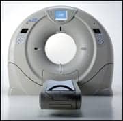
| The AquilionONE can image the entire heart in a single rotation, providing volumetric temporal resolution that is superior to multislice temporal resolution available today, resulting in clearer image quality. |
“MR makes much prettier pictures,” said Furqan Hussain Tejani, MD, FACC, director of advanced cardiovascular imaging at Long Island College Hospital, Brooklyn, NY. “It’s a much better acquisition modality and a much more sensitive and specific tool. Yet, it takes anywhere from 45 minutes to do an MR, and it only takes about 15 seconds to a minute to do cardiac CT. Any technique that has the capability of doing it faster is going to win the day.”
The noninvasive cardiac CT allows physicians to view the coronary arteries, particularly anomalies such as calcific plaque, noncalcific plaque, and narrowing of the arteries. “The real strength of cardiac CT is it has a high negative predictive value,” said David Rosenblum, DO, vice chairman of the department of radiology at Metro Health Medical Center, Cleveland. “It is excellent at ruling out coronary artery disease.”
Evolutionary Technology
Since its inception in the late 1970s, CT technology has evolved rapidly. Breakthroughs such as the move from single- to multidetector technology have paved the way to clearer images and better techniques to work around the motion of the heart. Today, advanced options include 64-slice, 128-slice, 256-slice, 320-slice, and dual-source models.
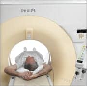 |
| The 256-slice Brilliance iCT scanner allows radiologists to produce high-quality images with exceptional acquisition speed, including complete coverage of the heart and brain. |
“With newer scanners—64-slice and above—you’re able to have thinner sections, which then reduces the distortion when you’re doing 3D imaging,” Rumberger said. “That allows you to look at those smaller structures, particularly the coronary arteries, more reliably.”
While the rotation speed is virtually the same for scanners with 64 slices and above, more slices mean faster scanning times. With a 64-slice detector, for example, it often takes three or four rotations to get a complete picture of the heart. “It’s like a jigsaw puzzle,” Rumberger said. “I’m putting four separate rotations into the computer and hoping that they line up.”
By contrast, Toshiba’s Aquilion ONE, which was launched at RSNA 2007, is a 320-slice scanner, allowing the entire heart (up to 16 cm of anatomy) to be imaged in one rotation. “If I take all of these slices at one time, there’s no jigsaw puzzle to put together,” Rumberger said. “So that helps the reliability of the imaging.”
Higher detector numbers also mean fewer respiratory motion artifacts. Metro Health Medical Center served as a beta-testing site for the 256-slice Brilliance iCT scanner from Philips. By performing six 180? rotations per second, the unit scans the heart within three heartbeats (5 to 8 seconds). “The beauty of it is the spatial resolution,” said Rosenblum, whose team also uses a 64-slice detector. “The ability to discriminate between two points is far greater with this scanner because it can resolve down to half a centimeter.”
Of course, CT has a long way to go to catch up with more invasive methods, such as catheterization. “The current resolution of CTA is about 0.4 mm, while for cardiac catheterization, it is 0.1 mm,” Rumberger said.
However, the implications for CT—especially on a diagnostic front—are vast. Coronary CTA can be used to confirm an open bypass graft, for example. According to Rosenblum, patients who present with acute coronary syndrome with low to intermediate risk of coronary artery disease would likely be ideal candidates for cardiac CT. Those who have uninterpretable EKGs or are unable to undergo a stress test could also benefit. He also sees potential for detecting ischemic cardiomyopathy in a noninvasive way.
 |
| The 256-slice Brilliance iCT scanner. |
CT scans could also be used to reveal coronary anomalies—an area that Rosenblum believes is better suited to CT than traditional catheterization. “There are certain malignant forms that go between the pulmonary artery and aorta that can be easily detected with coronary CTA,” he said.
On the other hand, CT is not ideal for patients with arrhythmias or for older patients who have densely calcified arteries. “If somebody has a lot of calcium, then it’s difficult to tell how much narrowing a vessel has with CT, even with the new machine,” Rosenblum said. He adds that CT is best for ruling out disease in younger patients, particularly those in their 30s or 40s, although those in their 50s and 60s without risk factors such as high blood pressure and diabetes would also benefit.
Dose Reduction
As more detectors have been included in modern 3D CT machines, the radiation dose has increased accordingly—a point of concern for many in the field. While Rumberger acknowledges that lowering the dose is ideal, he also cautions that the higher radiation exposure levels are often taken out of context.
“The radiation dose is higher than we would like it to be,” Rumberger said. “But if I did a 64-slice CTA on a patient, I would give the patient less radiation than he or she would get from a stress test with nuclear imaging.” He adds that the standard nuclear test requires twice the radiation of the CT scan.
He notes that manufacturers and researchers alike are working to minimize the dose of CT scans. “We all want to reduce the radiation dose just because it makes sense,” Rumberger said. “In fact, there are ways to reduce the dose significantly right now, and everybody practices that way.”
One popular reduction strategy is dose modulation, which involves turning on the scanner during only the part of the heart cycle that will provide the best motion-free images. Rosenblum notes that the Brilliance iCT scanner includes the “Step & Shoot” feature for this purpose. He also notes that the beam is more tightly “coned” than on the 64-slice detector, allowing for more precise targeting.
Tejani said that vendors continue to work on software and hardware algorithms to reduce the dose, and he is optimistic about their progress. “In the near future, we will arrive at a radiation exposure that will be low enough that you will not have the concern,” he said.
Usually, radiation concerns go hand-in-hand with concerns about increased use of contrast dyes—but not so with newer CT models. “Because the scanner is faster, you don’t need as much contrast,” Rosenblum said. “The boluses of contrast are smaller because you don’t need to image as long.”
Future Enhancements
There is no question that speed is one of cardiac CT’s most valuable assets. “You can’t really get much faster than what they’re getting now, so I think the speed and the coverage have really helped to free the structures so you can see them better,” Rosenblum said. “Anatomically, you’re more likely to get a good image than you were on the old scanners.”
As the technology continues to evolve, better viewing capabilities are expected. “Right now, we only have the capability to see the anatomy,” Tejani said. “We’re still trying to work our way through trying to see the physiology with CT.” The goal is to be able to assess profusion, an area that might involve the combined use of PET and CT.
Rosenblum also sees potential in using gating to detect arrhythmias in the heart, particularly those located in the pulmonary vein. “We used to just give patients a regular CT through the pulmonary vein,” he said. “Now, we’re using some gating to freeze the motion of the heart, and we get better images. Also, the thoracic aorta is better seen now in gating.”
Another area for improvement is in the display technology itself. Tejani pointed out the discrepancy between the 3D capabilities of the devices and traditional 2D monitors. Holographic displays would address that disconnect by allowing radiologists to see 3D images the way they are meant to be seen—in 3D space. “We will go from looking at the image to looking into the image,” Tejani said.
Most imaging suites are dark rooms, making them ideal for holographic displays. In the not too distant future, Tejani said 3D images will be projected from three cameras into a holographic box. He envisions images that will be sensitive to the touch, allowing radiologists to manipulate them in 3D space. “You actually will be able to hold the heart in your hand,” he said.
As the use of 3D CT becomes more widespread, the early detection capabilities of CT will become invaluable. Rumberger suggests that the technology will one day be a vital tool for primary care physicians to detect and treat heart disease in its earliest stages. He notes that current detection measures, such as measuring cholesterol levels, are unreliable. “Half of the people who come into the hospital with a heart attack have normal cholesterol,” he said. “It’s not high—it’s normal.”
Rumberger hopes that improved cardiac CT technology will encourage the health care profession to concentrate on ruling out blockages rather than focusing on stenosis. “The value of the 64-slice scanner is in saying, ?You do not have a blockage, but you are beginning to develop disease. And because you are beginning to develop disease—even though it’s not serious at this time—we can start making sense about treating you,’ ” Rumberger said.
Ann H. Carlson is a contributing writer for Medical Imaging. For more information, contact .

