 |
· HD7 Arrives in the US
· Toshiba Addresses Cardiac 4D Demand
· New Uses for MR-Guided Focused Ultrasound
HD7 Arrives in the US
Hi-def and affordable, too!

Royal Philips Electronics has unveiled a new high-definition ultrasound machine designed with the cost-conscious customer in mind. As a midrange ultrasound system, the Philips HD7 costs between $30,000 and $60,000 per unit, depending on the choice of transducers, said Jim R. Brown, senior director of clinical and technical marketing for Philips.
“The HD7 is good for the office environment, where their budgets might be limited but they still want to perform high-quality ultrasound,” Brown said.
The Philips HD7 boasts features that are more common on high-end ultrasound systems than midrange units, and the product is smaller and easier to move around than the more expensive models—which can cost up to $250,000, he said.
 |
| The HD7 from Philips is a high-definition ultrasound machine that offers tissue harmonic imaging capabilities. |
The high-definition system provides grayscale and color Doppler imaging. And the system can be used for anatomical M-Mode, a way to depict motion in a biological structure, such as the heart. The system has spectral Doppler and pulsatility index capabilities.
“It’s very easy to use, it’s very mobile,” Brown said. “Depending on the environment and what [customers] need to do, this product can be an absolutely perfect fit.”
The system can be used in a variety of high-volume settings, including cardiovascular, obstetrics/gynecology, anesthesiology, oncology, electrophysiology, stress echo, pediatric, orthopedic, urologic, and emergency applications.
The HD7’s competitors among midrange ultrasound systems include General Electric Healthcare’s LOGIQ P5 and LOGIC S6, as well as Siemens AG’s line of G model ultrasound machines. Philips in 2004 released its EnVisor, another midrange ultrasound system. The HD7 builds on some of the capabilities of the EnVisor.
“There is a very strong midrange market out there, so we would expect the HD7 to do as well as, if not better than, the EnVisor product,” Brown said. One of the big differences between the EnVisor and the HD7 is that the new system has an adjustable flat-panel LCD monitor, while the EnVisor relies on a cathode ray tube for displaying images, Brown said. And the control panel layout is more intuitive on the HD7 than on the EnVisor, he said.

Like the EnVisor, the HD7 has a feature called iSCAN that optimizes performance with the push of a button, such as by adjusting the brightness level of an image. The system makes those automatic adjustments based on built-in algorithms, Brown said. But the user also can override those settings with manual adjustments.
The HD7 also has a button to adjust to a patient’s size, allowing for optimal scanning of obese or small patients. And it has Tissue Harmonic Imaging, a mode that reduces unwanted artifacts by relying on the harmonic images instead of the fundamental frequency, Brown said. The HD7 relies on a broadband digital beamformer, as do all the other Philips ultrasound systems. With that technology, the system captures and preserves more tissue information than narrowband systems, according to Philips. Other features include multiple transducer ports, connectivity for digital image communication (DICOM), and easy data recording to CD or USB.
The HD7 was introduced to Europe in March, at the European Congress of Radiology in Vienna, and the product is just hitting the market in the United States. But Philips officials also have high hopes for the HD7 in the global market, because the product’s affordability makes it a good fit for hospitals and practices in the developing world, Brown said. “This product actually does very well in many areas of the world where they do have lots of budget constraints and they have to have a system that has multiple function capabilities and multiple application support,” Brown said.
—Alex Dobuzinskis
 |
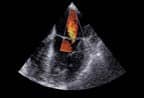
| Toshiba?s Aplio Artida features the ability to track myocardial motion. |
Toshiba Addresses Cardiac 4D Demand
Although the cardiac 4D market has been around for quite some time, it hadn’t been known for its efficiency—until recently, that is. Now, with a growing demand for real-time functionality, the big players in the industry are releasing solutions that aim to meet their customers’ needs.
The latest company to introduce one such product is Toshiba America Medical Systems Inc, Tustin, Calif, which in late March announced its brand-new Aplio Artida ultrasound system. According to Toshiba, it is the first ultrasound system in the world with the ability to track myocardial motion and display the images three-dimensionally.
The Artida offers real-time, multiplaner reformatting capabilities, which allow for the quantification of global and regional LV function. Bill Kenny, product manager, Ultrasound Business Unit, said, “The heart is three-dimensional, and now we can look at the whole heart in a cardiac cycle and perform a quantitative analysis.”
With the 2D/3D wall motion tracking features, users can obtain angle-independent, quantitative, and regional information about myocardial contraction, as well as optimize the pacemaker setting. Toshiba also anticipates that the ability to identify wall-motion defects and heart timing will tremendously improve Cardiac Resynchronization Therapy using pacemakers by examining whether a person will be a responder to CRT.
“Toshiba’s 4D tracking offers estimation of left ventricular volumes, ejection fraction, and direct measurement of global and regional myocardial function by means of speckle tracking, and provides insights that echocardiographers have not had before,” said Gerard Aurigemma, MD, professor of medicine at the University of Massachusetts and director of noninvasive cardiology at the UMass Memorial Medical Center.
Customer feedback played a huge role in bringing the Artida to market, Kenny said. Capture and propping tools allow for a rendered image in two steps, improving on other systems in the market that require 12 steps or more. In emphasizing ease of use, Toshiba also strived to reduce the development of repetitive stress syndrome in users, Kenny said.
The Artida is equipped with additional features that enhance diagnostic capabilities, such as multiple processors that run synchronously. This function gives way for the handling of large amounts of data, as well as heightens the level of imaging performance and clarity. Specifically, it uses the distributed processing power of more than 80 processor cores interconnected by a fast, digital system interface.
Advanced digital signal processing is accomplished through Artida’s SmartCast Beamformer, which precisely and flexibly controls the shape of the ultrasound. Also, with the product’s intuitive SmartSlice functions, physicians can cut, slice, and position the 4D volume quickly and conveniently. Other notable performance improvements consist of simultaneous multiple transmit focus or a doubled frame rate in color Doppler mode, helping to expedite exams while improving diagnostic accuracy.
Kenny stressed the importance of enhancing image quality to the fullest, a goal on which Toshiba is particular focused. “The better the image, the quicker the sonographer can capture the image, the quicker a physician can review the images, and the quicker the patient is completed,” he said.
—Elaine Sanchez
New Uses for MR-Guided Focused Ultrasound
In 2004, InSightec, a Dallas-based company owned by Elbit Imaging, General Electric, and MediTech Advisors, received approval from the FDA to treat women suffering from symptomatic uterine fibroids with its ExAblate MR-guided focused ultrasound surgery system.
Now looking beyond uterine fibroids, this year the company expects to start numerous pivotal oncology studies, including one investigating its effectiveness on brain tumors and another to test the system as a pain-relieving treatment for bone metastases.
Although the treatment is new to the health care area, it is fast becoming an alternative to conventional surgery. Neuroradiologist Suzanne LeBlang, MD, medical director, MR-Guided Focused Ultrasound Surgery, University MRI Diagnostic Treatment Center in Boca Raton, Fla, said her team has performed about 120 MR-guided focused ultrasound procedures since October 2004. She was introduced to the technology after application specialists from InSightec gave a lecture at her facility.
“What immediately impressed me was the ability to perform a ?surgical’ procedure without cutting the skin,” LeBlang said. “Also, it was amazing to see how the procedure was performed with complete image guidance and thermal monitoring of temperatures. This has never been done with this amount of precision, as MRI is the best anatomic imaging study available and can also act like a thermometer to follow temperature changes in the body as well.”
Using InSightec’s ExAblate 2000, with GE’s 1.5T and 3T MR systems, clinicians can employ a highly focused ultrasound in conjunction with an MR scanner to ablate fibroid tumors. The MR provides 3D images of the fibroid and surrounding tissue, ensuring precise guidance of ultrasound waves. It also enables the physician to determine real-time levels of heating and to monitor the progress of treatment during and after the procedure.
According to InSightec, the concept of focused ultrasound as a noninvasive treatment modality has been researched for more than 60 years, yet the lack of a good visualization system prevented it from taking off. In the 1990s, as a result of the widespread availability of ultrasound and MR, the outpatient, noninvasive procedure became a viable clinical option.
Recent studies have shown that with the ExAblate women sustained relief from uterine fibroid symptoms for up to 2 years, with a low incidence of side effects. Risks include skin burns, back or leg pain, abdominal cramping, nausea, fever, vaginal discharge, and urinary tract infection.
Physicians must examine the placement and size of fibroids to determine whether a patient is a candidate. LeBlang said that out of more than 250 patients scanned, she treats three to four cases per month. Procedure costs range from $10,000 to $15,000, and sites can work with health insurance companies to obtain preapproval coverage.
InSightec officials say the company is investing significant resources to develop application-specific software and hardware. Key applications it is targeting include pain relief for bone metastases and treatment for breast cancer, prostate cancer, brain tumors, and liver cancer. Future areas of research include the kidney, thyroid, pancreas, ovaries, and lymph nodes, to name a few, in addition to examining the possibility of treating central nervous diseases, like epilepsy and Parkinson’s, as well as targeted drug delivery.
—Elaine Sanchez

