A team of Johns Hopkins neurosurgeons and biomedical engineers has received $13.48 million from the Defense Advanced Research Projects Agency (DARPA) to develop implantable ultrasound and other devices that could revolutionize care for people suffering from spinal cord injuries. The results could benefit thousands of U.S. service members and civilians who sustain spinal cord injuries every year.
The electronic device is planned to be the size and flexibility of a small Band-Aid and will use high-resolution ultrasound technology to help doctors monitor and treat the changes in blood flow and prevent tissue death that occur immediately after a traumatic injury to the spinal cord.
The research program, supported by DARPA’s Bridging the Gap (BG+) program, will draw from the clinical expertise and ingenuity of its co-leaders, Nicholas Theodore, MD, professor of Neurosurgery and Biomedical Engineering and Amir Manbachi, PhD, assistant professor of Neurosurgery and Biomedical Engineering at the Johns Hopkins University School of Medicine, to bring the devices from concept to human use within an ambitious five-year timeline.
“There are few places taking a close look at how engineering approaches could improve the treatment of spinal cord injuries. I think there are tremendous opportunities here,” says Theodore, who also worked for more than 10 years as a neurosurgeon in the U.S. Navy, treating soldiers and sailors with spinal cord injuries.
Though the primary mission of the team is to develop devices that can be deployed to service members on military fronts, the researchers aim to make the technology available to benefit the approximately 17,000 civilians who experience spinal cord injuries in the United States every year.
“The main factors that make a device useable in a war theatre are size and ease of application in low-resource settings—both of which can only improve our clinical approaches as well,” says Theodore.
The project’s strategy is to target the disruption in blood flow that occurs alongside injury to the spinal cord. By utilizing technology to image and stimulate the blood vessels and tissue at the site of spinal cord injury, as well as controlling spinal fluid dynamics, the delivery of oxygen and nutrients can be optimized. This approach could prevent additional damage to the spinal cord, which can lead to increased inflammation, pain, and worsening paralysis.
“There are very few treatment options available to minimize the damage initially—for example, increasing the patient’s blood pressure; however, we still need to understand, in real time, how the body reacts to these treatments,” says Manbachi. “When Dr. Theodore described this challenge to me for the first time four years ago, as an acoustic engineer, I immediately thought to use ultrasound as a tool to monitor and stimulate these damaged tissues.”
To accomplish this, the electronic devices will use ultrasound “pulse echoes”—similar to the radar submarines use to navigate—as well as electrical stimulation, to monitor and treat the previously unobservable tiny blood vessels and surrounding tissue at and around the spinal cord injury site.
“This will be a real engineering feat,” says Manbachi. “Typical ultrasound transducers are bulky and designed to gather images of larger structures. We want to take this technology and shrink it for use on structures the size of a pinky finger, while still capturing clear ultrasound images of the spinal cord microvasculature.”
These images, say the researchers, will allow clinicians to observe how blood is flowing to the injury site. This can give them valuable information about how much oxygen, nutrients, and medication are reaching the area. The data will allow physicians to respond to their patient’s condition in real time by administering medications or possibly electrical or ultrasound stimulation to improve blood flow, stop inflammation, and offer pain relief and neuroprotective therapies to stop damage to the injured tissue.
The sound waves generated by these implantable devices can also be used to stimulate healing in the area. Similar to how the sun’s rays can be focused by a magnifying glass, therapeutic sound waves from the device can, in theory, be focused to promote blood flow at the injury site to promote healing.
The five-year timeline to bring a completely new technology to animal studies, FDA regulatory approvals, and human trials will push the limits of the typical pace of innovation that may otherwise take decades.
“It is an extraordinary team effort to bring together the smartest engineers and neurosurgeons to solve this problem,” says Theodore. “There are very few places in the world that are able to pull together the resources we have for this project.”
The team expects the initial technologies to be used experimentally and clinically to treat acute spinal cord injury. The researchers’ ultimate goal, however, is to develop more advanced versions applicable for patients suffering from chronic spinal cord injury.
The project will be coordinated by Senior Project Manager Chad Restrick, MSM-CRA, at Johns Hopkins University with key collaborators from the Johns Hopkins Medicine Departments of Neurosurgery, Biomedical Engineering, Radiology, Neurology and Critical Care Medicine, as well as external collaborators, including Ken Shepard, PhD, at Columbia University; Kyle Morrison, MS, of Sonic Concepts Inc.; Monique Beaudoin, PhD (Program Manager), George Coles, PhD, Francesco Tenore, PhD, and Steve Babin, MD-PhD (technical contributors) of the Johns Hopkins Applied Physics Laboratory.
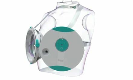
Featured image: Johns Hopkins scientists are developing an implantable ultrasound device to treat patients with traumatic spinal cord injuries. Credit: Ian Suk, Johns Hopkins