Little patients are receiving big care benefits from a variety of ultrasound technologies such as clearer 3D, revved-up echo, and transcranial Doppler imaging. The focus is on eliminating more invasive tests whenever necessary, assessing interventions, and simplifying workflow—all the while efficiently archiving these images for the baby book and, more important, the long-term medical archive.
No matter how you slice it—pediatric ultrasound is booming. New technologies and applications are sprouting up everywhere, resulting in better patient care for the pint-sized population and, at the same time, improving workflow for harried sonologists and clinicians. The advances encompass everything from high-end 3D systems to digital support tools, to new transducers and catheters. And ultrasound contrast is poised to make an even bigger splash in the not too distant future.
Advances in echocardiography such as Philips Medical Systems’ (Bothell, Wash) Live 3D Echo Technology and Siemens Medical Solutions’ (Malvern, Pa) AcuNav catheter are making it easier to observe and evaluate cardiac defects in adults and children. What about the oft-touted 3D ultrasound? Pediatric sonologists are just beginning to scratch its surface, but excitement is high. 3D and transcranial Doppler may be used in place of MRI or CT in certain situations. Zero radiation makes ultrasound especially appealing for pediatric use.
Like other imaging modalities, ultrasound is joining the march into the digital world, and radiologists are reporting significant gains in workflow courtesy of automated reporting systems. Real-time clips also have quite a bit of potential for freeing up radiologists; the technologist instead of the radiologist can complete the study, allowing the radiologist to remain in the department. Despite all of the buzz about new tools and applications, pediatric sonologists are clamoring for more. For example, higher-frequency transducers will fuel further applications of 3D. Sonologists also are eagerly awaiting the approval of a wider variety of contrast agents by the Food and Drug Administration (Washington, DC).
Pediatric Echo Roundup
Pediatric echocardiography may be the most challenging of all ultrasound imaging modalities. Patients’ rapid heart rates require higher frame rates and fast transition through modes. Moreover, resolution requirements are the greatest because the structures are the smallest and most complex. Finally, unlike adults, kids battle formative (versus degenerative) cardiac diseases. As a result, generic echocardiography solutions do not necessarily meet the needs of pediatric cardiologists. Several new technologies aim to make the modality a bit easier for pediatric cardiologists and their patients.
Norman Silverman, MD, professor of pediatric cardiology at Stanford University (Palo Alto, Calif), begins, “3D reconstruction looks like a very exciting prospect in echocardiography. It seems to be particularly useful at demonstrating complex valve abnormalities.” This detailed information, in turn, aids the surgical decision-making process.
One of the newest advances in the field is Philips’ Live 3D Echo technology. The technology offers a number of advantages; it is not only noninvasive but also provides exquisite images of the beating, and often tiny, heart. Girish Shirali, MD, associate professor of pediatrics and director of pediatric echocardiography at the Medical University of South Carolina (Charleston, SC), explains, “3D echo gives us the ability to noninvasively view the beating heart using a portable machine that can be wheeled to the patient’s bedside. Problem areas such as the mitral valve, ventricular septum, and left ventricular outflow tract are of particular interest since these are areas where 3D provides significantly more anatomic detail.”
The technology is especially beneficial for the pediatric population because it does not cause discomfort and eliminates the need for sedation. Moreover, once the image is captured, the clinician can manipulate it, which eliminates the need for multiple scans to capture different views of the heart and reduces costs.
In some cases, new tools are simplifying cardiac surgery. For example, Siemens’ AcuNav intracardiac ultrasound catheter can be used to observe cardiac structures and defects. Although the device is not approved for pediatric use, pediatric cardiologists have identified a few applications. Silverman explains, “Interventional cardiologists can insert the catheter through the neck or groin and use it in closures [of cardiac holes]. Because they are accustomed to using catheters, they can look at whatever they want without me there to guide them. It’s really revolutionized interventional procedures.” In fact, it is now possible to close some holes as a day procedure without anesthesia.
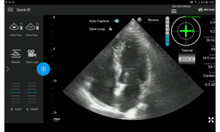
 Sonographer conducts a pediatric echocardiography study on the Acuson Sequoia system.
Pediatric cardiac patients may not be the only population that can benefit from advances in ultrasound catheters. In the future, fetuses may realize similar benefits. Researchers are using ultrasound to assist in fetal cardiac intervention by means of catheters. According to Silverman, it may be possible to help prevent progression of some forms of congenital heart disease, such as aortic stenosis, in the fetal stage. While fetal intervention is still in its infancy, early results are promising. The still-developing fetal heart shows incredible resilience in terms of tissue regeneration; postsurgical scarring is absolutely minimal. Results range from potentially improved outcomes to relieved obstructions.
Achieving such results is no small feat and will require a change in mind-set. “If disease is preventable during fetal development, primary obstetrical sonographers must complete careful early evaluation,” Silverman says. Typically, most of the information can be obtained via a four-chamber view of the heart with an extension of that view during the 18th to 25th week of gestation.
Finally, tissue Doppler ultrasound shows some promise in pediatric cardiology. The tissue Doppler method can be used to measure and describe diastolic function in children, thus providing surgeons with information to assess the timing of interventions for kids. Another option that is currently under investigation is Strain Rate Imaging (SRI). SRI is an extension of tissue Doppler that measures the rate of tissue deformation and can differentiate between actively deforming and passive muscle segments to provide information on left ventricular flow.
All the Dimensions
3D is here, it’s here to stay, and it is associated with a number of advantages. Lynn Ansley Fordham, MD, chief of pediatric imaging and associate professor at the University of North Carolina School of Medicine (Chapel Hill, NC), says, “3D scanning allows experienced and less experienced examiners to get equally good results.” For example, with a 3D system, sonologists can reorient or straighten a slightly crooked scan. Fordham and her colleagues use a Medison (Cypress, Calif) Voluson 3D system for cranial ultrasounds of premature infants. With a 3D ultrasound system, sonologists can get the entire volume in a single acquisition; they can see the entire brain in one view instead of multiple slices. Fordham explains, “We have more information much more quickly.” Achieving comparable results with a routine 2D scan of the head requires three separate scans. In other scenarios, such as abdominal scans, it is nearly impossible to obtain a third view with a routine scan.
Although 3D does yield volumes of data and additional views, Fordham reports that it does not negatively impact workflow. In fact, she says, once users learn how to use the machine and workstation, 3D saves time because the sonologist does not have to rescan as much.
At this point, pediatric sonologists are merely scratching the surface of the potential of 3D. Fordham says, “I’m hoping to use it for liver transplant work and routine renal studies, but I’d like to see higher-frequency transducers made available first.” Higher-frequency transducers result in better image resolution, which is essential for Fordham’s tiny customers.
Of course, budgets are always a concern with any new technology purchase. Yet, the utility of 3D could work in its favor. Fordham explains, “3D is an exciting technology that will yield useful information for a variety of applications.” That useful information can, in turn, help fund an investment in a 3D system. Case in point? Currently, pediatric liver transplant patients must undergo a CT scan, complete with radiation, or a higher-priced MRI scan prior to surgery. 3D ultrasound, however, can yield the data in a single, quick acquisition. And unlike CT or MRI, ultrasound typically does not entail sedation.
Still, not all 3D systems are created equal. Budget-conscious facilities may be intrigued by the price tag of 3D handheld systems, and these systems can certainly enhance the accessibility and portability of 3D. The end results, however, may not offer much additional information in terms of clinical utility. Fordham says, “Using a high-end system with motorized transducers allows for uniform image acquisition, reliable reconstruction, and accurate volume measurement.” On the other hand, less expensive systems generally lack motorized transducers. The upshot? “They may create beautiful pictures, but you can’t make a volume measurement,” says Fordham.
New and Not So New
3D does not hold a monopoly on eliminating sedation and radiation for pediatric patients. David Blews, MD, president of Children’s Diagnostic Imaging of Atlanta PC (Atlanta), practices at Children’s Healthcare of Atlanta (Atlanta) and is completing a research project using transcranial Doppler to measure intracerebral circulation in patients with sickle-cell anemia to predict which patients have the highest risk of stroke and offer preventive treatment to patients at high risk. Blews says, “It’s very promising. This is rapidly becoming the standard of care.” The only other ways to obtain similar information about sickle-cell patients’ risk of stroke is via an MR angiogram or a conventional angiogram. Neither of these tests is ideal; an MR angiogram is expensive and requires sedation, and a conventional angiogram is invasive. Blews adds, “Transcranial Doppler picks up velocity changes earlier than the anatomical changes detected with the other tests. In addition to having an earlier predictive value, transcranial Doppler is noninvasive and less expensive, and does not require sedation.”
Some older techniques, including extended field of view (EFOV) ultrasound, are being rediscovered and reinvigorating the modality. Brian Coley, MD, chief of the section of ultrasound and assistant director of the department of radiology at Columbus Children’s Hospital (Columbus, Ohio), explains, “Sometimes clinicians have difficulty with ultrasound. It does not have the same global view as CT or MRI.” EFOV ultrasound can provide those all-important landmarks, which can assist radiologists as well as clinicians. Coley and his colleagues use EFOV for neonatal spine imaging to check for a tethered cord. He explains, “EFOV allows us to paint a broad view of the spine and lets us count better.” A static view, however, forces sonologists to count as they scan. Another EFOV application is tumor imaging; EFOV can show clinicians the full extent of the tumor.
While EFOV provides that broad, big-picture view, real-time clips can also be used to orient physicians and sonologists. Coley says, “The technologist can sweep through the region and give you a sense of being there.” One advantage of real time is on the all-important labor end. The technologist can complete a portable study in the ICU, while the radiologist remains in the department. In fact, some sonologists are beginning to debate the pros and cons of completing a certain number of still pictures versus real-time sweeps. The answer remains up in the air and may vary according to the needs at individual settings. Regardless of the answer, some sonologists will always prefer to personally scan the patient. Blews of Children’s Diagnostic Imaging of Atlanta opines, “There’s no substitute for doing your own scanning for true sonologists. We find that being there is really key.”
Another application of real-time clips is remote imaging in rural settings. Coley explains, “Real-time clips let you do a quick, remote exam and link to a radiologist through a PAC system.” The radiologist can assess the patient remotely and possibly eliminate an unnecessary trip to the urban hospital.
In some cases, ultrasound does not eliminate other tests. Instead, it is used in conjunction with other techniques, proving the old adage that the sum of the whole is greater than its parts. Blews is using fluoroscopy and ultrasound to do image-guided injections of joint spaces. How does it work? He injects the patient with gadolinium under the guidance of fluoroscopy and ultrasound and then completes an MR arthrogram. “It’s useful for a wide variety of pediatric joint disorders and in trauma cases. The combination allows us to detect very subtle abnormalities of the tendons and cartilage that could not otherwise be detected by MRI.”
Workflow, Workflow, Workflow
Let’s face it. It is wonderful to have a new 3D system at your disposal, and it is great to find new applications for older technologies, but sometimes what really drives physicians and administrators are the tools that simplify workflow. The pediatric echo lab is no exception. In fact, it faces a downright information overload. So it is no surprise that Silverman claims his biggest cause for excitement is the support tools for reading ultrasound studies in children.
Until recently, pediatric cardiology required videotape recording of ultrasound studies. The results are enormous libraries of stored videotape. Silverman notes, “It requires a tedious amount of time to read through all of these.” Unfortunately, time is an all too precious commodity in pediatric cardiology.
Cardiologists at Stanford seem to have found a solution. Silverman explains, “We are moving toward a total digital environment, where all information is centralized by means of a computer server.” Short clips are stored (and easily recalled) on the server. A pediatric automated reporting system simplifies the reporting process. The physician selects statements about parameters such as the size and location of a defect from a menu of choices. The next step is to modify and add to each statement as necessary. The system interfaces with the hospital’s PACS and is automatically connected to its backbone. The results? It no longer takes 8 to 10 days for the most recent report to appear on the hospital system; it is immediately available. The system also allows cardiologists to maximize their time and view studies remotely. They also can look at studies from other sites. Silverman concludes, “It provides a great opportunity to centralize and make the practice more efficient.”
On the Horizon
More than a few pediatric sonologists are eagerly waiting the more widespread availability and use of ultrasound contrast agents in the United States. Ultrasound contrast is widely used in the rest of the world, but Coley predicts it is probably 1 or 2 years away from increased use here. Still that has not stopped pediatric sonologists from considering the applications.The premise of ultrasound contrast is quite simple. Most contrast agents are used to increase Doppler signals. The short list of pediatric applications is quite impressive. Contrast could be used to distinguish benign from malignant liver masses and potentially obviate the need for a biopsy. It also could improve signal strength for transcranial Doppler scans, providing a clear image of intracranial vessels. Contrast-enhanced scans could replace fluoroscopy as the test of choice to look for reflux in kids with recurrent urinary tract infections. The primary advantage is, once again, eliminating radiation.
“Once we get contrast,” says Coley, “people will find uses for it.” In fact, given the recent concerns about pediatric radiation exposure, there may be some demand from patients’ parents to use ultrasound instead of other imaging procedures in certain cases.
In the meantime, pediatric sonologists and their patients are reaping a variety of benefits from new technologies. Patient care is improved via higher-quality images in 3D and echo. Costs can be reduced in many cases courtesy of fewer rescans and replacement of higher-priced imaging procedures. Finally, the digital revolution has reached sonology, and the result is enhanced workflow.