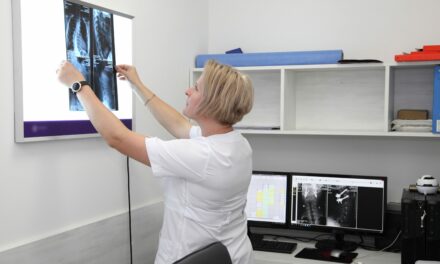
Eastern Daily Press profiles Tom Turmezei, MD, PhD, consultant radiologist at Norfolk and Norwich University Hospital in the United Kingdom, who discusses the role of 3D imaging in clinical care.
“We can identify at-risk patients, but x-rays aren’t always sensitive or accurate enough for us to definitively tell a patient that they’re at risk of a joint replacement within a certain amount of years because of specific factors.
“It was through everyday practice that about 12 years ago I started to think: ‘There must be a better way of looking at this’.
“The technique we’re developing looks at the whole joint in 3D, so it shows things that x-rays can’t. We use a CT (computer tomography) scanner which is very good at looking at structures containing minerals like calcium, like you find in bones. Then we use a computer algorithm that takes measurements of the joint much more reliably and accurately than a human could.”
Read more at Eastern Daily Press.