Scientists observe real-time dopamine release during demanding cognitive tasks, potentially paving the way for targeted therapies in conditions like Parkinson’s and ADHD.
Scientists have confirmed a neurobiochemical link between dopamine and cognitive flexibility, according to new research published in the March issue of The Journal of Nuclear Medicine.
PET imaging shows that the brain increases dopamine production when completing cognitively demanding tasks, and that the more dopamine released, the more efficiently the tasks are completed. Armed with this information, physicians may soon be able to develop more precise treatment strategies for neurological and psychiatric disorders.
Cognitive flexibility is the ability to adapt one’s thinking and behavior appropriately to a changing environment and is considered an aspect of executive function. Cognitive flexibility differs among people and is reported to be impaired in several psychiatric and neurologic disorders, such as depression, posttraumatic stress disorder, addiction, anxiety disorder, schizophrenia, Parkinson’s disease, and attention-deficit/hyperactivity disorder.
“At the neurotransmitter level, the dopamine system has been linked to cognitive flexibility. A direct neurochemical response to cognitive flexibility, however, has yet to be shown,” says Isabelle Miederer, PhD, associate professor in experimental nuclear medicine in the department of nuclear medicine at University Medical Center Mainz, Germany, in a release. “In our study, we sought to examine the release of dopamine in real-time by performing PET scans while individuals completed behavioral flexibility tasks.”
Study Methodology and Findings
Eighteen participants were scanned with the D2/3 receptor ligand 18F-fallypride in a two-part block study design. In the first part, participants performed two tasks consecutively on a computer screen without rule switching while undergoing PET imaging. In the second part of the PET scan, participants had to switch flexibly between two task rules. Dopamine release was calculated using the linearized simplified reference region model which compares the two task blocks with each other.
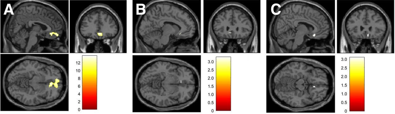
PET imaging analysis showed a displacement of 18F-fallypride in the ventromedial prefrontal cortex during the task switching (higher cognitive demand) part of the study, which is assumed to be the release of dopamine. Results also showed that the greater dopamine release, the more efficient participants were in switching between tasks.
“The present findings emphasize the significance of dopamine in cognitive flexibility,” says Mathias Schreckenberger, MD, head of the department of nuclear medicine at University Medical Center Mainz, in a release. “They are consistent with the results of previous clinical studies indicating that dopamine deficiency in disorders such as Parkinson’s disease may cause behavioral deficits in cognitive flexibility.
“Looking forward, it is expected that the results of the study will contribute to a better understanding of the neurochemical mechanisms underlying cognitive flexibility and thus facilitate the development of treatment strategies to improve flexibility in neurological and psychiatric disorders.”
ID 68224615 © Sudok1 | Dreamstime.com